Click image to see more details
-

-
-
-
-
+6
Product Info Summary
| SKU: | M08821-2 |
|---|---|
| Size: | 100 μg/vial |
| Reactive Species: | Human, Mouse |
| Host: | Mouse |
| Application: | Flow Cytometry, IHC, WB |
Customers Who Bought This Also Bought

Product info
Product Name
Anti-Histone H1.0/H1F0 Antibody Picoband® (monoclonal, 5I3E6)
SKU/Catalog Number
M08821-2
Size
100 μg/vial
Form
Lyophilized
Description
Boster Bio Anti-Histone H1.0/H1F0 Antibody Picoband® (monoclonal, 5I3E6) catalog # M08821-2. Tested in Flow Cytometry, IHC, WB applications. This antibody reacts with Human, Mouse. The brand Picoband indicates this is a premium antibody that guarantees superior quality, high affinity, and strong signals with minimal background in Western blot applications. Only our best-performing antibodies are designated as Picoband, ensuring unmatched performance.
Storage & Handling
At -20°C for one year from date of receipt. After reconstitution, at 4°C for one month. It can also be aliquotted and stored frozen at -20°C for six months. Avoid repeated freezing and thawing.
Cite This Product
Anti-Histone H1.0/H1F0 Antibody Picoband® (monoclonal, 5I3E6) (Boster Biological Technology, Pleasanton CA, USA, Catalog # M08821-2)
Host
Mouse
Contents
Each vial contains 4 mg Trehalose, 0.9 mg NaCl and 0.2 mg Na2HPO4.
Clonality
Monoclonal
Clone Number
5I3E6
Isotype
Mouse IgG2b
Immunogen
E.coli-derived human Histone H1.0/H1F0 recombinant protein (Position: K20-K159).
*Blocking peptide can be purchased. Costs vary based on immunogen length. Contact us for pricing.
Cross-reactivity
No cross-reactivity with other proteins.
Reactive Species
M08821-2 is reactive to H1F0 in Human, Mouse
Reconstitution
Adding 0.2 ml of distilled water will yield a concentration of 500 μg/ml.
Observed Molecular Weight
24 kDa
Calculated molecular weight
20.863kDa
Background of H1-0
H1 histone family, member 0is a member of thehistonefamily of nuclearproteinswhich are a component ofchromatin. In humans, this protein is encoded by theH1F0gene. It is mapped to 22q13.1. Histones are basic nuclear proteins that are responsible for the nucleosome structure of the chromosomal fiber in eukaryotes. Nucleosomes consist of approximately 146 bp of DNA wrapped around a histone octamer composed of pairs of each of the four core histones (H2A, H2B, H3, and H4). The chromatin fiber is further compacted through the interaction of a linker histone, H1, with the DNA between the nucleosomes to form higher order chromatin structures. This gene is intronless and encodes a replication-independent histone that is a member of the histone H1 family.
Antibody Validation
Boster validates all antibodies on WB, IHC, ICC, Immunofluorescence, and ELISA with known positive control and negative samples to ensure specificity and high affinity, including thorough antibody incubations.
Application & Images
Applications
M08821-2 is guaranteed for Flow Cytometry, IHC, WB Boster Guarantee
Assay Dilutions Recommendation
The recommendations below provide a starting point for assay optimization. The actual working concentration varies and should be decided by the user.
Western blot, 0.25-0.5 μg/ml, Human
Immunohistochemistry(Paraffin-embedded Section), 2-5 μg/ml, Human, Mouse
Flow Cytometry (Fixed), 1-3 μg/1x106 cells, Human
Positive Control
WB: human U20S whole cell, human PC-3 whole cell, human HepG2 whole cell
IHC: mouse colon tissue, human placenta tissue, human differentiated adenocarcinoma of the rectum tissue, human Hodgkin's lymphoma tissue, human renal cell carcinoma tissue, human tonsil tissue, human gastric carcinoma tissue, human liver cancer tissue
FCM: SiHa cell
Validation Images & Assay Conditions
Click image to see more details
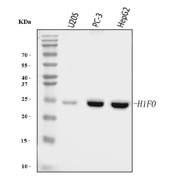
Figure 1. Western blot analysis of Histone H1.0/H1F0 using anti-Histone H1.0/H1F0 antibody (M08821-2).
Electrophoresis was performed on a 5-20% SDS-PAGE gel at 70V (Stacking gel) / 90V (Resolving gel) for 2-3 hours. The sample well of each lane was loaded with 30 ug of sample under reducing conditions.
Lane 1: human U20S whole cell lysates,
Lane 2: human PC-3 whole cell lysates,
Lane 3: human HepG2 whole cell lysates.
After electrophoresis, proteins were transferred to a nitrocellulose membrane at 150 mA for 50-90 minutes. Blocked the membrane with 5% non-fat milk/TBS for 1.5 hour at RT. The membrane was incubated with mouse anti-Histone H1.0/H1F0 antigen affinity purified monoclonal antibody (Catalog # M08821-2) at 0.5 μg/mL overnight at 4°C, then washed with TBS-0.1%Tween 3 times with 5 minutes each and probed with a goat anti-mouse IgG-HRP secondary antibody at a dilution of 1:10000 for 1.5 hour at RT. The signal is developed using an Enhanced Chemiluminescent detection (ECL) kit (Catalog # EK1001) with Tanon 5200 system. A specific band was detected for Histone H1.0/H1F0 at approximately 24 kDa. The expected band size for Histone H1.0/H1F0 is at 24 kDa.

Click image to see more details
Figure 2. IHC analysis of Histone H1.0/H1F0 using anti-Histone H1.0/H1F0 antibody (M08821-2).
Histone H1.0/H1F0 was detected in a paraffin-embedded section of mouse colon tissue. Heat mediated antigen retrieval was performed in EDTA buffer (pH 8.0, epitope retrieval solution). The tissue section was blocked with 10% goat serum. The tissue section was then incubated with 2 μg/ml mouse anti-Histone H1.0/H1F0 Antibody (M08821-2) overnight at 4°C. Peroxidase Conjugated Goat Anti-mouse IgG was used as secondary antibody and incubated for 30 minutes at 37°C. The tissue section was developed using HRP Conjugated Mouse IgG Super Vision Assay Kit (Catalog # SV0001) with DAB as the chromogen.

Click image to see more details
Figure 3. IHC analysis of Histone H1.0/H1F0 using anti-Histone H1.0/H1F0 antibody (M08821-2).
Histone H1.0/H1F0 was detected in a paraffin-embedded section of human placenta tissue. Heat mediated antigen retrieval was performed in EDTA buffer (pH 8.0, epitope retrieval solution). The tissue section was blocked with 10% goat serum. The tissue section was then incubated with 2 μg/ml mouse anti-Histone H1.0/H1F0 Antibody (M08821-2) overnight at 4°C. Peroxidase Conjugated Goat Anti-mouse IgG was used as secondary antibody and incubated for 30 minutes at 37°C. The tissue section was developed using HRP Conjugated Mouse IgG Super Vision Assay Kit (Catalog # SV0001) with DAB as the chromogen.

Click image to see more details
Figure 4. IHC analysis of Histone H1.0/H1F0 using anti-Histone H1.0/H1F0 antibody (M08821-2).
Histone H1.0/H1F0 was detected in a paraffin-embedded section of human differentiated adenocarcinoma of the rectum tissue. Heat mediated antigen retrieval was performed in EDTA buffer (pH 8.0, epitope retrieval solution). The tissue section was blocked with 10% goat serum. The tissue section was then incubated with 2 μg/ml mouse anti-Histone H1.0/H1F0 Antibody (M08821-2) overnight at 4°C. Peroxidase Conjugated Goat Anti-mouse IgG was used as secondary antibody and incubated for 30 minutes at 37°C. The tissue section was developed using HRP Conjugated Mouse IgG Super Vision Assay Kit (Catalog # SV0001) with DAB as the chromogen.

Click image to see more details
Figure 5. IHC analysis of Histone H1.0/H1F0 using anti-Histone H1.0/H1F0 antibody (M08821-2).
Histone H1.0/H1F0 was detected in a paraffin-embedded section of human Hodgkin's lymphoma tissue. Heat mediated antigen retrieval was performed in EDTA buffer (pH 8.0, epitope retrieval solution). The tissue section was blocked with 10% goat serum. The tissue section was then incubated with 2 μg/ml mouse anti-Histone H1.0/H1F0 Antibody (M08821-2) overnight at 4°C. Peroxidase Conjugated Goat Anti-mouse IgG was used as secondary antibody and incubated for 30 minutes at 37°C. The tissue section was developed using HRP Conjugated Mouse IgG Super Vision Assay Kit (Catalog # SV0001) with DAB as the chromogen.

Click image to see more details
Figure 6. IHC analysis of Histone H1.0/H1F0 using anti-Histone H1.0/H1F0 antibody (M08821-2).
Histone H1.0/H1F0 was detected in a paraffin-embedded section of human renal cell carcinoma tissue. Heat mediated antigen retrieval was performed in EDTA buffer (pH 8.0, epitope retrieval solution). The tissue section was blocked with 10% goat serum. The tissue section was then incubated with 2 μg/ml mouse anti-Histone H1.0/H1F0 Antibody (M08821-2) overnight at 4°C. Peroxidase Conjugated Goat Anti-mouse IgG was used as secondary antibody and incubated for 30 minutes at 37°C. The tissue section was developed using HRP Conjugated Mouse IgG Super Vision Assay Kit (Catalog # SV0001) with DAB as the chromogen.

Click image to see more details
Figure 7. IHC analysis of Histone H1.0/H1F0 using anti-Histone H1.0/H1F0 antibody (M08821-2).
Histone H1.0/H1F0 was detected in a paraffin-embedded section of human tonsil tissue. Heat mediated antigen retrieval was performed in EDTA buffer (pH 8.0, epitope retrieval solution). The tissue section was blocked with 10% goat serum. The tissue section was then incubated with 2 μg/ml mouse anti-Histone H1.0/H1F0 Antibody (M08821-2) overnight at 4°C. Peroxidase Conjugated Goat Anti-mouse IgG was used as secondary antibody and incubated for 30 minutes at 37°C. The tissue section was developed using HRP Conjugated Mouse IgG Super Vision Assay Kit (Catalog # SV0001) with DAB as the chromogen.

Click image to see more details
Figure 8. IHC analysis of Histone H1.0/H1F0 using anti-Histone H1.0/H1F0 antibody (M08821-2).
Histone H1.0/H1F0 was detected in a paraffin-embedded section of human gastric carcinoma tissue. Heat mediated antigen retrieval was performed in EDTA buffer (pH 8.0, epitope retrieval solution). The tissue section was blocked with 10% goat serum. The tissue section was then incubated with 2 μg/ml mouse anti-Histone H1.0/H1F0 Antibody (M08821-2) overnight at 4°C. Peroxidase Conjugated Goat Anti-mouse IgG was used as secondary antibody and incubated for 30 minutes at 37°C. The tissue section was developed using HRP Conjugated Mouse IgG Super Vision Assay Kit (Catalog # SV0001) with DAB as the chromogen.

Click image to see more details
Figure 9. IHC analysis of Histone H1.0/H1F0 using anti-Histone H1.0/H1F0 antibody (M08821-2).
Histone H1.0/H1F0 was detected in a paraffin-embedded section of human liver cancer tissue. Heat mediated antigen retrieval was performed in EDTA buffer (pH 8.0, epitope retrieval solution). The tissue section was blocked with 10% goat serum. The tissue section was then incubated with 2 μg/ml mouse anti-Histone H1.0/H1F0 Antibody (M08821-2) overnight at 4°C. Peroxidase Conjugated Goat Anti-mouse IgG was used as secondary antibody and incubated for 30 minutes at 37°C. The tissue section was developed using HRP Conjugated Mouse IgG Super Vision Assay Kit (Catalog # SV0001) with DAB as the chromogen.

Click image to see more details
Figure 10. Flow Cytometry analysis of SiHa cells using anti-Histone H1.0/H1F0 antibody (M08821-2).
Overlay histogram showing SiHa cells stained with M08821-2 (Blue line). To facilitate intracellular staining, cells were fixed with 4% paraformaldehyde and permeabilized with permeabilization buffer. The cells were blocked with 10% normal goat serum. And then incubated with mouse anti-Histone H1.0/H1F0 Antibody (M08821-2, 1 μg/1x106 cells) for 30 min at 20°C. DyLight®488 conjugated goat anti-mouse IgG (BA1126, 5-10 μg/1x106 cells) was used as secondary antibody for 30 minutes at 20°C. Isotype control antibody (Green line) was mouse IgG (1 μg/1x106) used under the same conditions. Unlabelled sample without incubation with primary antibody and secondary antibody (Red line) was used as a blank control.
Protein Target Info & Infographic
Gene/Protein Information For H1F0 (Source: Uniprot.org, NCBI)
Gene Name
H1F0
Full Name
Histone H1.0
Weight
20.863kDa
Superfamily
histone H1/H5 family
Alternative Names
T-complex protein 1 subunit gamma; TCP-1-gamma; CCT-gamma; hTRiC5; CCT3; CCTG; TRIC5 H1-0 H1.0, H10, H1F0, H1FV H1.0 linker histone histone H1.0|H1 histone family member 0|H1.0, H1(0), H1-0|histone H1|histone H1(0)
*If product is indicated to react with multiple species, protein info is based on the gene entry specified above in "Species".For more info on H1F0, check out the H1F0 Infographic

We have 30,000+ of these available, one for each gene! Check them out.
In this infographic, you will see the following information for H1F0: database IDs, superfamily, protein function, synonyms, molecular weight, chromosomal locations, tissues of expression, subcellular locations, post-translational modifications, and related diseases, research areas & pathways. If you want to see more information included, or would like to contribute to it and be acknowledged, please contact [email protected].
Specific Publications For Anti-Histone H1.0/H1F0 Antibody Picoband® (monoclonal, 5I3E6) (M08821-2)
Hello CJ!
No publications found for M08821-2
*Do you have publications using this product? Share with us and receive a reward. Ask us for more details.
Recommended Resources
Here are featured tools and databases that you might find useful.
- Boster's Pathways Library
- Protein Databases
- Bioscience Research Protocol Resources
- Data Processing & Analysis Software
- Photo Editing Software
- Scientific Literature Resources
- Research Paper Management Tools
- Molecular Biology Software
- Primer Design Tools
- Bioinformatics Tools
- Phylogenetic Tree Analysis
Customer Reviews
Have you used Anti-Histone H1.0/H1F0 Antibody Picoband® (monoclonal, 5I3E6)?
Submit a review and receive an Amazon gift card.
- $30 for a review with an image
0 Reviews For Anti-Histone H1.0/H1F0 Antibody Picoband® (monoclonal, 5I3E6)
Customer Q&As
Have a question?
Find answers in Q&As, reviews.
Can't find your answer?
Submit your question
