Click image to see more details
Product Info Summary
| SKU: | A01876-1 |
|---|---|
| Size: | 100 μg/vial |
| Reactive Species: | Human, Mouse |
| Host: | Rabbit |
| Application: | ELISA, IHC, WB |
Customers Who Bought This Also Bought


Product info
Product Name
Anti-Histidine decarboxylase/HDC Antibody Picoband®
SKU/Catalog Number
A01876-1
Size
100 μg/vial
Form
Lyophilized
Description
Boster Bio Anti-Histidine decarboxylase/HDC Antibody Picoband® catalog # A01876-1. Tested in ELISA, IHC, WB applications. This antibody reacts with Human, Mouse. The brand Picoband indicates this is a premium antibody that guarantees superior quality, high affinity, and strong signals with minimal background in Western blot applications. Only our best-performing antibodies are designated as Picoband, ensuring unmatched performance.
Storage & Handling
At -20°C for one year from date of receipt. After reconstitution, at 4°C for one month. It can also be aliquotted and stored frozen at -20°C for six months. Avoid repeated freezing and thawing.
Cite This Product
Anti-Histidine decarboxylase/HDC Antibody Picoband® (Boster Biological Technology, Pleasanton CA, USA, Catalog # A01876-1)
Host
Rabbit
Contents
Each vial contains 4 mg Trehalose, 0.9 mg NaCl, 0.2 mg Na2HPO4.
Clonality
Polyclonal
Isotype
Rabbit IgG
Immunogen
E.coli-derived human Histidine decarboxylase/HDC recombinant protein (Position: R12-V662).
*Blocking peptide can be purchased. Costs vary based on immunogen length. Contact us for pricing.
Cross-reactivity
No cross-reactivity with other proteins.
Reactive Species
A01876-1 is reactive to HDC in Human, Mouse
Reconstitution
Adding 0.2 ml of distilled water will yield a concentration of 500 μg/ml.
Observed Molecular Weight
74 kDa
Calculated molecular weight
50433 MW
Background of HDC
HDC(Histidine decarboxylase) is the enzyme that catalyzes the reaction that produces histamine from histidine with the help of vitamin B6. The HDC gene is mapped on 15q21.2. The deduced 662-amino acid protein has a molecular mass of 74,148 Da. The gene contains 12 exons spanning approximately 24 kb. The biogenic amine histamine is an important modulator of numerous physiologic processes, including neurotransmission, gastric acid secretion, and smooth muscle tone. The biosynthesis of histamine from histidine is catalyzed by the enzyme L-histidine decarboxylase. This homodimeric enzyme is a pyridoxal phosphate(PLP)-dependent decarboxylase and is highly specific for its histidine substrate. Ercan-Sencicek et al. noted that animal studies had shown that lack of Hdc in mice results in increased locomotor and stereotypic behaviors, as well as increased anxiety.
Antibody Validation
Boster validates all antibodies on WB, IHC, ICC, Immunofluorescence, and ELISA with known positive control and negative samples to ensure specificity and high affinity, including thorough antibody incubations.
Application & Images
Applications
A01876-1 is guaranteed for ELISA, IHC, WB Boster Guarantee
Assay Dilutions Recommendation
The recommendations below provide a starting point for assay optimization. The actual working concentration varies and should be decided by the user.
Western blot, 0.25-0.5 μg/ml, Human
Immunohistochemistry(Paraffin-embedded Section), 2-5 μg/ml, Mouse
ELISA, 0.1-0.5 μg/ml, Human
Positive Control
WB: human HEL whole cell, human K562 whole cell, human Hacat whole cell, human HL-60 whole cell
IHC: mouse brain tissue
Validation Images & Assay Conditions
Click image to see more details
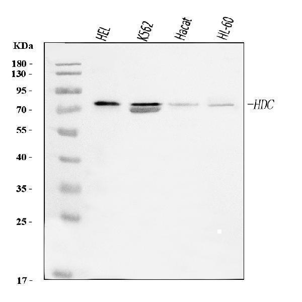
Figure 1. Western blot analysis of Histidine Decarboxylase/HDC using anti-Histidine Decarboxylase/HDC antibody (A01876-1).
Electrophoresis was performed on a 5-20% SDS-PAGE gel at 70V (Stacking gel) / 90V (Resolving gel) for 2-3 hours. The sample well of each lane was loaded with 30 ug of sample under reducing conditions.
Lane 1: human HEL whole cell lysates,
Lane 2: human K562 whole cell lysates,
Lane 3: human Hacat whole cell lysates,
Lane 4: human HL-60 whole cell lysates.
After electrophoresis, proteins were transferred to a nitrocellulose membrane at 150 mA for 50-90 minutes. Blocked the membrane with 5% non-fat milk/TBS for 1.5 hour at RT. The membrane was incubated with rabbit anti-Histidine Decarboxylase/HDC antigen affinity purified polyclonal antibody (Catalog # A01876-1) at 0.5 μg/mL overnight at 4°C, then washed with TBS-0.1%Tween 3 times with 5 minutes each and probed with a goat anti-rabbit IgG-HRP secondary antibody at a dilution of 1:5000 for 1.5 hour at RT. The signal is developed using an Enhanced Chemiluminescent detection (ECL) kit (Catalog # EK1002) with Tanon 5200 system. A specific band was detected for Histidine Decarboxylase/HDC at approximately 74 kDa. The expected band size for Histidine Decarboxylase/HDC is at 74 kDa.

Click image to see more details
Figure 2. IHC analysis of Histidine Decarboxylase/HDC using anti-Histidine Decarboxylase/HDC antibody (A01876-1).
Histidine Decarboxylase/HDC was detected in a paraffin-embedded section of mouse brain tissue. Heat mediated antigen retrieval was performed in EDTA buffer (pH 8.0, epitope retrieval solution). The tissue section was blocked with 10% goat serum. The tissue section was then incubated with 2 μg/ml rabbit anti-Histidine Decarboxylase/HDC Antibody (A01876-1) overnight at 4°C. Peroxidase Conjugated Goat Anti-rabbit IgG was used as secondary antibody and incubated for 30 minutes at 37°C. The tissue section was developed using HRP Conjugated Rabbit IgG Super Vision Assay Kit (Catalog # SV0002) with DAB as the chromogen.
Protein Target Info & Infographic
Gene/Protein Information For HDC (Source: Uniprot.org, NCBI)
Gene Name
HDC
Full Name
Histidine decarboxylase
Weight
50433 MW
Superfamily
group II decarboxylase family
Alternative Names
Neuromodulin; Axonal membrane protein GAP-43; Growth-associated protein 43; Neural phosphoprotein B-50; pp46; GAP43 HDC histidine decarboxylase histidine decarboxylase
*If product is indicated to react with multiple species, protein info is based on the gene entry specified above in "Species".For more info on HDC, check out the HDC Infographic

We have 30,000+ of these available, one for each gene! Check them out.
In this infographic, you will see the following information for HDC: database IDs, superfamily, protein function, synonyms, molecular weight, chromosomal locations, tissues of expression, subcellular locations, post-translational modifications, and related diseases, research areas & pathways. If you want to see more information included, or would like to contribute to it and be acknowledged, please contact [email protected].
Specific Publications For Anti-Histidine decarboxylase/HDC Antibody Picoband® (A01876-1)
Hello CJ!
No publications found for A01876-1
*Do you have publications using this product? Share with us and receive a reward. Ask us for more details.
Recommended Resources
Here are featured tools and databases that you might find useful.
- Boster's Pathways Library
- Protein Databases
- Bioscience Research Protocol Resources
- Data Processing & Analysis Software
- Photo Editing Software
- Scientific Literature Resources
- Research Paper Management Tools
- Molecular Biology Software
- Primer Design Tools
- Bioinformatics Tools
- Phylogenetic Tree Analysis
Customer Reviews
Have you used Anti-Histidine decarboxylase/HDC Antibody Picoband®?
Submit a review and receive an Amazon gift card.
- $30 for a review with an image
0 Reviews For Anti-Histidine decarboxylase/HDC Antibody Picoband®
Customer Q&As
Have a question?
Find answers in Q&As, reviews.
Can't find your answer?
Submit your question
