Click image to see more details
Product Info Summary
| SKU: | A00839 |
|---|---|
| Size: | 100 μg/vial |
| Reactive Species: | Human, Mouse, Rat |
| Host: | Rabbit |
| Application: | ELISA, Flow Cytometry, WB |
Customers Who Bought This Also Bought

Product info
Product Name
Anti-HDAC3 Antibody Picoband®
SKU/Catalog Number
A00839
Size
100 μg/vial
Form
Lyophilized
Description
Boster Bio Anti-HDAC3 Antibody Picoband® catalog # A00839. Tested in ELISA, Flow Cytometry, WB applications. This antibody reacts with Human, Mouse, Rat. The brand Picoband indicates this is a premium antibody that guarantees superior quality, high affinity, and strong signals with minimal background in Western blot applications. Only our best-performing antibodies are designated as Picoband, ensuring unmatched performance.
Storage & Handling
Store at -20˚C for one year from date of receipt. After reconstitution, at 4˚C for one month. It can also be aliquotted and stored frozen at -20˚C for six months. Avoid repeated freeze-thaw cycles.
Cite This Product
Anti-HDAC3 Antibody Picoband® (Boster Biological Technology, Pleasanton CA, USA, Catalog # A00839)
Host
Rabbit
Contents
Each vial contains 4 mg Trehalose, 0.9 mg NaCl and 0.2 mg Na2HPO4.
Clonality
Polyclonal
Isotype
Rabbit IgG
Immunogen
E.coli-derived human HDAC3 recombinant protein (Position: M1-I428).
*Blocking peptide can be purchased. Costs vary based on immunogen length. Contact us for pricing.
Cross-reactivity
No cross-reactivity with other proteins.
Reactive Species
A00839 is reactive to HDAC3 in Human, Mouse, Rat
Reconstitution
Add 0.2ml of distilled water will yield a concentration of 500ug/ml.
Observed Molecular Weight
49 kDa
Calculated molecular weight
55914 MW
Background of HDAC3
HDAC3 (HISTONE DEACETYLASE 3) is a member of the histone deacetylase/acuc/apha family of proteins that is an enzyme that in humans is encoded by the HDAC3 gene. The HDAC3 gene is mapped to 5q31.3. HDAC3 has histone deacetylase activity and represses transcription when tethered to a promoter. It may participate in the regulation of transcription through its binding with the zinc-finger transcription factor YY1. The protein can also down-regulate p53 function and thus modulate cell growth and apoptosis. And this gene is regarded as a potential tumor suppressor gene. HDAC3 has an open reading frame of 428 amino acids and shares 53% amino acid identity with HDAC1 and 52% with HDAC2. The catalytic domain of HDAC4 interacts with HDAC3 via the transcriptional corepressor NCOR2. All experimental conditions leading to the suppression of HDAC4 binding to NCOR2 and to HDAC3 resulted in loss of enzymatic activity associated with HDAC4. HDAC3 recruitment to the genome displays a circadian rhythm in mouse liver.
Antibody Validation
Boster validates all antibodies on WB, IHC, ICC, Immunofluorescence, and ELISA with known positive control and negative samples to ensure specificity and high affinity, including thorough antibody incubations.
Application & Images
Applications
A00839 is guaranteed for ELISA, Flow Cytometry, WB Boster Guarantee
Assay Dilutions Recommendation
The recommendations below provide a starting point for assay optimization. The actual working concentration varies and should be decided by the user.
Western blot, 0.1-0.5μg/ml
Flow Cytometry (Fixed), 1-3 μg/1x106 cells
ELISA, 0.1-0.5μg/ml
Positive Control
WB: human 293T whole cell, human Hela whole cell, human HepG2 whole cell, human MOLT-4 whole cell, human MCF-7 whole cell, human K562 whole cell, human CACO-2 whole cell, rat thymus tissue, rat C6 whole cell, mouse ANA-1 whole cell, mouse NIH/3T3 whole cell
FCM: MCF-7 cell, RAW2647 cell, RH35 cell
Validation Images & Assay Conditions
Click image to see more details
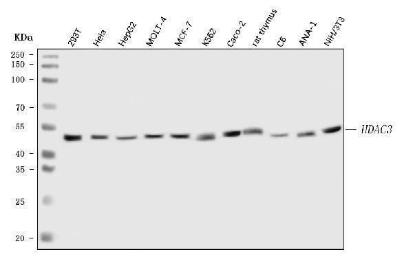
Figure 1. Western blot analysis of HDAC3 using anti-HDAC3 antibody (A00839).
Electrophoresis was performed on a 5-20% SDS-PAGE gel at 70V (Stacking gel) / 90V (Resolving gel) for 2-3 hours. The sample well of each lane was loaded with 30 ug of sample under reducing conditions.
Lane 1: human 293T whole cell lysates,
Lane 2: human Hela whole cell lysates,
Lane 3: human HepG2 whole cell lysates,
Lane 4: human MOLT-4 whole cell lysates,
Lane 5: human MCF-7 whole cell lysates,
Lane 6: human K562 whole cell lysates,
Lane 7: human CACO-2 whole cell lysates,
Lane 8: rat thymus tissue lysates,
Lane 9: rat C6 whole cell lysates,
Lane 10: mouse ANA-1 whole cell lysates,
Lane 11: mouse NIH/3T3 whole cell lysates.
After electrophoresis, proteins were transferred to a nitrocellulose membrane at 150 mA for 50-90 minutes. Blocked the membrane with 5% non-fat milk/TBS for 1.5 hour at RT. The membrane was incubated with rabbit anti-HDAC3 antigen affinity purified polyclonal antibody (Catalog # A00839) at 0.5 μg/mL overnight at 4°C, then washed with TBS-0.1%Tween 3 times with 5 minutes each and probed with a goat anti-rabbit IgG-HRP secondary antibody at a dilution of 1:5000 for 1.5 hour at RT. The signal is developed using an Enhanced Chemiluminescent detection (ECL) kit (Catalog # EK1002) with Tanon 5200 system. A specific band was detected for HDAC3 at approximately 49 kDa. The expected band size for HDAC3 is at 49 kDa.

Click image to see more details
Figure 2. Flow Cytometry analysis of MCF-7 cells using anti-HDAC3 antibody (A00839).
Overlay histogram showing MCF-7 cells stained with A00839 (Blue line). To facilitate intracellular staining, cells were fixed with 4% paraformaldehyde and permeabilized with permeabilization buffer. The cells were blocked with 10% normal goat serum. And then incubated with rabbit anti-HDAC3 Antibody (A00839, 1 μg/1x106 cells) for 30 min at 20°C. DyLight®488 conjugated goat anti-rabbit IgG (BA1127, 5-10 μg/1x106 cells) was used as secondary antibody for 30 minutes at 20°C. Isotype control antibody (Green line) was rabbit IgG (1 μg/1x106) used under the same conditions. Unlabelled sample without incubation with primary antibody and secondary antibody (Red line) was used as a blank control.

Click image to see more details
Figure 3. Flow Cytometry analysis of RAW264.7 cells using anti-HDAC3 antibody (A00839).
Overlay histogram showing RAW264.7 cells stained with A00839 (Blue line). To facilitate intracellular staining, cells were fixed with 4% paraformaldehyde and permeabilized with permeabilization buffer. The cells were blocked with 10% normal goat serum. And then incubated with rabbit anti-HDAC3 Antibody (A00839, 1 μg/1x106 cells) for 30 min at 20°C. DyLight®488 conjugated goat anti-rabbit IgG (BA1127, 5-10 μg/1x106 cells) was used as secondary antibody for 30 minutes at 20°C. Isotype control antibody (Green line) was rabbit IgG (1 μg/1x106) used under the same conditions. Unlabelled sample without incubation with primary antibody and secondary antibody (Red line) was used as a blank control.

Click image to see more details
Figure 4. Flow Cytometry analysis of RH35 cells using anti-HDAC3 antibody (A00839).
Overlay histogram showing RH35 cells stained with A00839 (Blue line). To facilitate intracellular staining, cells were fixed with 4% paraformaldehyde and permeabilized with permeabilization buffer. The cells were blocked with 10% normal goat serum. And then incubated with rabbit anti-HDAC3 Antibody (A00839, 1 μg/1x106 cells) for 30 min at 20°C. DyLight®488 conjugated goat anti-rabbit IgG (BA1127, 5-10 μg/1x106 cells) was used as secondary antibody for 30 minutes at 20°C. Isotype control antibody (Green line) was rabbit IgG (1 μg/1x106) used under the same conditions. Unlabelled sample without incubation with primary antibody and secondary antibody (Red line) was used as a blank control.
Protein Target Info & Infographic
Gene/Protein Information For HDAC3 (Source: Uniprot.org, NCBI)
Gene Name
HDAC3
Full Name
Histone deacetylase 3
Weight
55914 MW
Superfamily
histone deacetylase family
Alternative Names
Histone deacetylase 3; HD3; RPD3-2; SMAP45; HDAC3 HDAC3 HD3, KDAC3, RPD3, RPD3-2 histone deacetylase 3 histone deacetylase 3|SMAP45
*If product is indicated to react with multiple species, protein info is based on the gene entry specified above in "Species".For more info on HDAC3, check out the HDAC3 Infographic

We have 30,000+ of these available, one for each gene! Check them out.
In this infographic, you will see the following information for HDAC3: database IDs, superfamily, protein function, synonyms, molecular weight, chromosomal locations, tissues of expression, subcellular locations, post-translational modifications, and related diseases, research areas & pathways. If you want to see more information included, or would like to contribute to it and be acknowledged, please contact [email protected].
Specific Publications For Anti-HDAC3 Antibody Picoband® (A00839)
Hello CJ!
A00839 has been cited in 1 publications:
*The publications in this section are manually curated by our staff scientists. They may differ from Bioz's machine gathered results. Both are accurate. If you find a publication citing this product but is missing from this list, please let us know we will issue you a thank-you coupon.
Interleukin-18 down-regulates multidrug resistance-associated protein 2 expression through farnesoid x receptor associated with nuclear factor kappa B and %u2026
Recommended Resources
Here are featured tools and databases that you might find useful.
- Boster's Pathways Library
- Protein Databases
- Bioscience Research Protocol Resources
- Data Processing & Analysis Software
- Photo Editing Software
- Scientific Literature Resources
- Research Paper Management Tools
- Molecular Biology Software
- Primer Design Tools
- Bioinformatics Tools
- Phylogenetic Tree Analysis
Customer Reviews
Have you used Anti-HDAC3 Antibody Picoband®?
Submit a review and receive an Amazon gift card.
- $30 for a review with an image
0 Reviews For Anti-HDAC3 Antibody Picoband®
Customer Q&As
Have a question?
Find answers in Q&As, reviews.
Can't find your answer?
Submit your question
16 Customer Q&As for Anti-HDAC3 Antibody Picoband®
Question
Is a blocking peptide available for product anti-HDAC3 antibody (A00839)?
Verified Customer
Verified customer
Asked: 2020-04-08
Answer
We do provide the blocking peptide for product anti-HDAC3 antibody (A00839). If you would like to place an order for it please contact [email protected] and make a special request.
Boster Scientific Support
Answered: 2020-04-08
Question
Would A00839 anti-HDAC3 antibody work on parafin embedded sections? If so, which fixation method do you recommend we use (PFA, paraformaldehyde, other)?
Verified Customer
Verified customer
Asked: 2020-03-05
Answer
You can see on the product datasheet, A00839 anti-HDAC3 antibody as been validated on WB. It is best to use PFA for fixation because it has better tissue penetration ability. PFA needs to be prepared fresh before use. Long term stored PFA turns into formalin, as the PFA molecules congregate and become formalin.
Boster Scientific Support
Answered: 2020-03-05
Question
I was wanting to use your anti-HDAC3 antibody for WB for rat skin on frozen tissues, but I want to know if it has been tested for this particular application. Has this antibody been tested and is this antibody a good choice for rat skin identification?
Verified Customer
Verified customer
Asked: 2020-01-09
Answer
It shows on the product datasheet, A00839 anti-HDAC3 antibody has been validated for ELISA, WB on human, mouse, rat tissues. We have an innovator award program that if you test this antibody and show it works in rat skin in IHC-frozen, you can get your next antibody for free.
Boster Scientific Support
Answered: 2020-01-09
Question
I have a question about product A00839, anti-HDAC3 antibody. I was wondering if it would be possible to conjugate this antibody with biotin. I would need it to be without BSA or sodium azide. I am planning on using a buffer exchange of sodium azide with PBS only. Would there be problems for me to conjugate the antibody and store it in -20 degrees in small aliquots?
Verified Customer
Verified customer
Asked: 2019-12-18
Answer
It is not recommended storing this antibody with PBS buffer only in -20 degrees. If you want to store it in -20 degrees it is best to add some cryoprotectant like glycerol. If you want carrier free A00839 anti-HDAC3 antibody, we can provide it to you in a special formula with trehalose and/or glycerol. These molecules will not interfere with conjugation chemistry and provide a good level of protection for the antibody from degradation. Please be sure to specify this in your purchase order.
Boster Scientific Support
Answered: 2019-12-18
Question
Will anti-HDAC3 antibody A00839 work for WB with skin?
Verified Customer
Verified customer
Asked: 2019-12-16
Answer
According to the expression profile of skin, HDAC3 is highly expressed in skin. So, it is likely that anti-HDAC3 antibody A00839 will work for WB with skin.
Boster Scientific Support
Answered: 2019-12-16
Question
We have tried in the past anti-HDAC3 antibody for WB on right hemisphere of cerebellum a few years ago. I am using rat, and We intend to use the antibody for ELISA next. I was wanting to use examining right hemisphere of cerebellum as well as leukemic t-cell in our next experiment. Could you please give me some suggestion on which antibody would work the best for ELISA?
M. Yang
Verified customer
Asked: 2019-11-28
Answer
I have checked the website and datasheets of our anti-HDAC3 antibody and I see that A00839 has been validated on rat in both WB and ELISA. Thus A00839 should work for your application. Our Boster satisfaction guarantee will cover this product for ELISA in rat even if the specific tissue type has not been validated. We do have a comprehensive range of products for ELISA detection and you can check out our website bosterbio.com to find out more information about them.
Boster Scientific Support
Answered: 2019-11-28
Question
Our team were well pleased with the WB result of your anti-HDAC3 antibody. However we have observed positive staining in erythroleukemia nucleus. using this antibody. Is that expected? Could you tell me where is HDAC3 supposed to be expressed?
Verified Customer
Verified customer
Asked: 2019-10-30
Answer
From literature, erythroleukemia does express HDAC3. Generally HDAC3 expresses in nucleus. Regarding which tissues have HDAC3 expression, here are a few articles citing expression in various tissues:
Cervix carcinoma, Pubmed ID: 17081983
Erythroleukemia, Pubmed ID: 23186163
Fibroblast, Pubmed ID: 9346952
Leukemic T-cell, Pubmed ID: 19690332
Skin, Pubmed ID: 15489334
Spleen, and T-cell, Pubmed ID: 9464271
Boster Scientific Support
Answered: 2019-10-30
Question
We appreciate helping with my inquiry over the phone. Here are the WB image, lot number and protocol we used for skin using anti-HDAC3 antibody A00839. Let me know if you need anything else.
Verified Customer
Verified customer
Asked: 2019-09-25
Answer
Thank you for the data. You have provided everything we needed. Our lab team are working to resolve your inquiry as quickly as possible, and we appreciate your patience and understanding! Please let me know if there is anything you need in the meantime.
Boster Scientific Support
Answered: 2019-09-25
Question
I am looking for using your anti-HDAC3 antibody for p75ntr negatively regulates cell cycle via sc1 studies. Has this antibody been tested with western blotting on a431 whole cell lysates? We would like to see some validation images before ordering.
Verified Customer
Verified customer
Asked: 2019-08-06
Answer
Thank you for your inquiry. This A00839 anti-HDAC3 antibody is tested on human k562, k562 whole cell lysates, a431 whole cell lysates. It is guaranteed to work for ELISA, WB in human, mouse, rat. Our Boster guarantee will cover your intended experiment even if the sample type has not been be directly tested.
Boster Scientific Support
Answered: 2019-08-06
Question
Here is the WB image, lot number and protocol we used for skin using anti-HDAC3 antibody A00839. Please let me know if you require anything else.
Verified Customer
Verified customer
Asked: 2018-09-27
Answer
Thank you very much for the data. Our lab team are working to resolve this as quickly as possible, and we appreciate your patience and understanding! You have provided everything we needed. Please let me know if there is anything you need in the meantime.
Boster Scientific Support
Answered: 2018-09-27
Question
I see that the anti-HDAC3 antibody A00839 works with WB, what is the protocol used to produce the result images on the product page?
Verified Customer
Verified customer
Asked: 2018-01-24
Answer
You can find protocols for WB on the "support/technical resources" section of our navigation menu. If you have any further questions, please send an email to [email protected]
Boster Scientific Support
Answered: 2018-01-24
Question
We have observed staining in mouse leukemic t-cell. Are there any suggestions? Is anti-HDAC3 antibody supposed to stain leukemic t-cell positively?
S. Yang
Verified customer
Asked: 2017-10-17
Answer
From what I have seen in literature leukemic t-cell does express HDAC3. From what I have seen in Uniprot.org, HDAC3 is expressed in right hemisphere of cerebellum, spleen t-cell, fibroblast, skin, cervix carcinoma, leukemic t-cell, erythroleukemia, among other tissues. Regarding which tissues have HDAC3 expression, here are a few articles citing expression in various tissues:
Cervix carcinoma, Pubmed ID: 17081983
Erythroleukemia, Pubmed ID: 23186163
Fibroblast, Pubmed ID: 9346952
Leukemic T-cell, Pubmed ID: 19690332
Skin, Pubmed ID: 15489334
Spleen, and T-cell, Pubmed ID: 9464271
Boster Scientific Support
Answered: 2017-10-17
Question
Is this A00839 anti-HDAC3 antibody reactive to the isotypes of HDAC3?
S. Wu
Verified customer
Asked: 2017-08-16
Answer
The immunogen of A00839 anti-HDAC3 antibody is E.coli-derived human HDAC3 recombinant protein (Position: M1-I428). Could you tell me which isotype you are interested in so I can help see if the immunogen is part of this isotype?
Boster Scientific Support
Answered: 2017-08-16
Question
I would like to test anti-HDAC3 antibody A00839 on rat skin for research purposes, then I may be interested in using anti-HDAC3 antibody A00839 for diagnostic purposes as well. Is the antibody suitable for diagnostic purposes?
E. Huang
Verified customer
Asked: 2017-08-07
Answer
The products we sell, including anti-HDAC3 antibody A00839, are only intended for research use. They would not be suitable for use in diagnostic work. If you have the means to develop a product into diagnostic use, and are interested in collaborating with us and develop our product into an IVD product, please contact us for more discussions.
Boster Scientific Support
Answered: 2017-08-07
Question
We are currently using anti-HDAC3 antibody A00839 for human tissue, and we are content with the ELISA results. The species of reactivity given in the datasheet says human, mouse, rat. Is it likely that the antibody can work on dog tissues as well?
M. Li
Verified customer
Asked: 2015-12-16
Answer
The anti-HDAC3 antibody (A00839) has not been validated for cross reactivity specifically with dog tissues, but there is a good chance of cross reactivity. We have an innovator award program that if you test this antibody and show it works in dog you can get your next antibody for free. Please contact me if I can help you with anything.
Boster Scientific Support
Answered: 2015-12-16
Question
Do you have a BSA free version of anti-HDAC3 antibody A00839 available?
R. Yang
Verified customer
Asked: 2015-02-04
Answer
I appreciate your recent telephone inquiry. I can confirm that some lots of this anti-HDAC3 antibody A00839 are BSA free. For now, these lots are available and we can make a BSA free formula for you free of charge. It will take 3 extra days to prepare. If you require this antibody BSA free again in future, please do not hesitate to contact me and I will be pleased to check which lots we have in stock that are BSA free.
Boster Scientific Support
Answered: 2015-02-04
