Click image to see more details
-

-
-
-
-
+3
Product Info Summary
| SKU: | M02561-2 |
|---|---|
| Size: | 100 μg/vial |
| Reactive Species: | Human, Mouse, Rat |
| Host: | Mouse |
| Application: | Flow Cytometry, IF, IHC, ICC, WB |
Customers Who Bought This Also Bought

Product info
Product Name
Anti-Grp75 Antibody Picoband® (monoclonal, 4I9)
View all GRP75/HSPA9B/Mortalin Antibodies
SKU/Catalog Number
M02561-2
Size
100 μg/vial
Form
Lyophilized
Description
Boster Bio Anti-Grp75 Antibody Picoband® (monoclonal, 4I9) catalog # M02561-2. Tested in Flow Cytometry, IF, IHC, ICC, WB applications. This antibody reacts with Human, Mouse, Rat. The brand Picoband indicates this is a premium antibody that guarantees superior quality, high affinity, and strong signals with minimal background in Western blot applications. Only our best-performing antibodies are designated as Picoband, ensuring unmatched performance.
Storage & Handling
At -20°C for one year from date of receipt. After reconstitution, at 4°C for one month. It can also be aliquotted and stored frozen at -20°C for six months. Avoid repeated freezing and thawing.
Cite This Product
Anti-Grp75 Antibody Picoband® (monoclonal, 4I9) (Boster Biological Technology, Pleasanton CA, USA, Catalog # M02561-2)
Host
Mouse
Contents
Each vial contains 4 mg Trehalose, 0.9 mg NaCl and 0.2 mg Na2HPO4.
Clonality
Monoclonal
Clone Number
4I9
Isotype
Mouse IgG1
Immunogen
A synthetic peptide corresponding to a sequence at the C-terminus of human Grp75, identical to the related mouse and rat sequences.
*Blocking peptide can be purchased. Costs vary based on immunogen length. Contact us for pricing.
Cross-reactivity
No cross-reactivity with other proteins.
Reactive Species
M02561-2 is reactive to HSPA9 in Human, Mouse, Rat
Reconstitution
Adding 0.2 ml of distilled water will yield a concentration of 500 μg/ml.
Observed Molecular Weight
74 kDa
Calculated molecular weight
73.68kDa
Background of GRP75/HSPA9B/Mortalin
HSPA9 (heat shock 70kDa protein 9 (mortalin)), also known as GRP75, mot-2, mthsp75, PBP74, HSPA9B, MORTALIN or MORTALIN, PERINUCLEAR, is a highly conserved member of the HSP70 family of proteins. It functions as a chaperone in the mitochondria, cytoplasm, and centrosome. The HSPA9 gene is mapped to chromosome 5q31.2 based on an alignment of the HSPA9 sequence with the genomic sequence. Knockdown of HSPA9 in erythroid cultures was associated with an increased number of cells in the G0/G1 phase of the cell cycle and accelerated apoptosis. Knockdown of Hspa9 in mouse bone marrow cells, followed by transplantation into wildtype recipients, also resulted in loss of erythroid cell number. Haploinsufficiency for HSPA9 may contribute to abnormal hematopoiesis in myelodysplastic syndromes. This protein plays a role in the control of cell proliferation.
Antibody Validation
Boster validates all antibodies on WB, IHC, ICC, Immunofluorescence, and ELISA with known positive control and negative samples to ensure specificity and high affinity, including thorough antibody incubations.
Application & Images
Applications
M02561-2 is guaranteed for Flow Cytometry, IF, IHC, ICC, WB Boster Guarantee
Assay Dilutions Recommendation
The recommendations below provide a starting point for assay optimization. The actual working concentration varies and should be decided by the user.
Western blot, 0.25-0.5 μg/ml, Human, Mouse, Rat
Immunohistochemistry(Paraffin-embedded Section), 2-5 μg/ml, Human
Immunocytochemistry/Immunofluorescence, 5 μg/ml, Human
Flow Cytometry (Fixed), 1-3 μg/1x106 cells, Human
Positive Control
WB: human Hela whole cell, human HepG2 whole cell, rat brain tissue, rat lung tissue, rat PC-12 whole cell, mouse brain tissue, mouse lung tissue, mouse RAW2647 whole cell
IHC: human appendiceal adenocarcinoma tissue, human gastric cancer tissue, human gall bladder adenosquamous carcinoma tissue, human lymphadenoma tissue
ICC/IF: CACO-2 cell
FCM: HepG2 cell
Validation Images & Assay Conditions
Click image to see more details
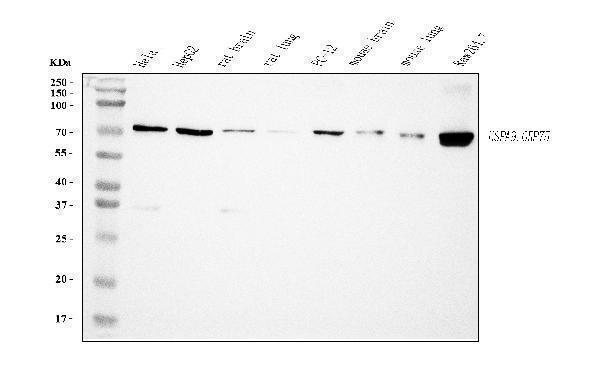
Figure 1. Western blot analysis of Grp75 using anti-Grp75 antibody (M02561-2).
Electrophoresis was performed on a 5-20% SDS-PAGE gel at 70V (Stacking gel) / 90V (Resolving gel) for 2-3 hours. The sample well of each lane was loaded with 30 ug of sample under reducing conditions.
Lane 1: human Hela whole cell lysates,
Lane 2: human HepG2 whole cell lysates,
Lane 3: rat brain tissue lysates,
Lane 4: rat lung tissue lysates,
Lane 5: rat PC-12 whole cell lysates,
Lane 6: mouse brain tissue lysates,
Lane 7: mouse lung tissue lysates,
Lane 8: mouse RAW264.7 whole cell lysates.
After electrophoresis, proteins were transferred to a nitrocellulose membrane at 150 mA for 50-90 minutes. Blocked the membrane with 5% non-fat milk/TBS for 1.5 hour at RT. The membrane was incubated with mouse anti-Grp75 antigen affinity purified monoclonal antibody (Catalog # M02561-2) at 0.5 μg/mL overnight at 4°C, then washed with TBS-0.1%Tween 3 times with 5 minutes each and probed with a goat anti-mouse IgG-HRP secondary antibody at a dilution of 1:10000 for 1.5 hour at RT. The signal is developed using an Enhanced Chemiluminescent detection (ECL) kit (Catalog # EK1001) with Tanon 5200 system. A specific band was detected for Grp75 at approximately 74 kDa. The expected band size for Grp75 is at 74 kDa.

Click image to see more details
Figure 2. IHC analysis of Grp75 using anti-Grp75 antibody (M02561-2).
Grp75 was detected in a paraffin-embedded section of human appendiceal adenocarcinoma tissue. Heat mediated antigen retrieval was performed in EDTA buffer (pH 8.0, epitope retrieval solution). The tissue section was blocked with 10% goat serum. The tissue section was then incubated with 2 μg/ml mouse anti-Grp75 Antibody (M02561-2) overnight at 4°C. Biotinylated goat anti-mouse IgG was used as secondary antibody and incubated for 30 minutes at 37°C. The tissue section was developed using Strepavidin-Biotin-Complex (SABC) (Catalog # SA1021) with DAB as the chromogen.

Click image to see more details
Figure 3. IHC analysis of Grp75 using anti-Grp75 antibody (M02561-2).
Grp75 was detected in a paraffin-embedded section of human gastric cancer tissue. Heat mediated antigen retrieval was performed in EDTA buffer (pH 8.0, epitope retrieval solution). The tissue section was blocked with 10% goat serum. The tissue section was then incubated with 2 μg/ml mouse anti-Grp75 Antibody (M02561-2) overnight at 4°C. Biotinylated goat anti-mouse IgG was used as secondary antibody and incubated for 30 minutes at 37°C. The tissue section was developed using Strepavidin-Biotin-Complex (SABC) (Catalog # SA1021) with DAB as the chromogen.

Click image to see more details
Figure 4. IHC analysis of Grp75 using anti-Grp75 antibody (M02561-2).
Grp75 was detected in a paraffin-embedded section of human gall bladder adenosquamous carcinoma tissue. Heat mediated antigen retrieval was performed in EDTA buffer (pH 8.0, epitope retrieval solution). The tissue section was blocked with 10% goat serum. The tissue section was then incubated with 2 μg/ml mouse anti-Grp75 Antibody (M02561-2) overnight at 4°C. Biotinylated goat anti-mouse IgG was used as secondary antibody and incubated for 30 minutes at 37°C. The tissue section was developed using Strepavidin-Biotin-Complex (SABC) (Catalog # SA1021) with DAB as the chromogen.

Click image to see more details
Figure 5. IHC analysis of Grp75 using anti-Grp75 antibody (M02561-2).
Grp75 was detected in a paraffin-embedded section of human lymphadenoma tissue. Heat mediated antigen retrieval was performed in EDTA buffer (pH 8.0, epitope retrieval solution). The tissue section was blocked with 10% goat serum. The tissue section was then incubated with 2 μg/ml mouse anti-Grp75 Antibody (M02561-2) overnight at 4°C. Biotinylated goat anti-mouse IgG was used as secondary antibody and incubated for 30 minutes at 37°C. The tissue section was developed using Strepavidin-Biotin-Complex (SABC) (Catalog # SA1021) with DAB as the chromogen.

Click image to see more details
Figure 6. IF analysis of Grp75 using anti-Grp75 antibody (M02561-2).
Grp75 was detected in an immunocytochemical section of CACO-2 cells. Enzyme antigen retrieval was performed using IHC enzyme antigen retrieval reagent (AR0022) for 15 mins. The cells were blocked with 10% goat serum. And then incubated with 5 μg/mL mouse anti-Grp75 Antibody (M02561-2) overnight at 4°C. DyLight®488 Conjugated Goat Anti-Mouse IgG (BA1126) was used as secondary antibody at 1:100 dilution and incubated for 30 minutes at 37°C. The section was counterstained with DAPI. Visualize using a fluorescence microscope and filter sets appropriate for the label used.

Click image to see more details
Figure 7. Flow Cytometry analysis of HepG2 cells using anti-Grp75 antibody (M02561-2).
Overlay histogram showing HepG2 cells stained with M02561-2 (Blue line). To facilitate intracellular staining, cells were fixed with 4% paraformaldehyde and permeabilized with permeabilization buffer. The cells were blocked with 10% normal goat serum. And then incubated with mouse anti-Grp75 Antibody (M02561-2, 1 μg/1x106 cells) for 30 min at 20°C. DyLight®488 conjugated goat anti-mouse IgG (BA1126, 5-10 μg/1x106 cells) was used as secondary antibody for 30 minutes at 20°C. Isotype control antibody (Green line) was mouse IgG (1 μg/1x106) used under the same conditions. Unlabelled sample without incubation with primary antibody and secondary antibody (Red line) was used as a blank control.
Protein Target Info & Infographic
Gene/Protein Information For HSPA9 (Source: Uniprot.org, NCBI)
Gene Name
HSPA9
Full Name
Stress-70 protein, mitochondrial
Weight
73.68kDa
Superfamily
heat shock protein 70 family
Alternative Names
CMT2L; CRYAC; DHMN 2; DHMN2; E2IG1; H11; HMN 2; HMN2; HMN2A; HSB8; HSPB 8; HspB8 HSPA9 CRP40, CSA, EVPLS, GRP-75, GRP75, HEL-S-124mB, MOT, MOT2, MTHSP75, PBP74, SAAN, SIDBA4, HSPA9 heat shock protein family A (Hsp70) member 9 stress-70 protein, mitochondrial|75 kDa glucose-regulated protein|catecholamine-regulated protein 40|epididymis secretory sperm binding protein Li 124m|heat shock 70kD protein 9B|heat shock 70kDa protein 9 (mortalin)|mortalin, perinuclear|mortalin-2|mortalin2|p66-mortalin|peptide-binding protein 74
*If product is indicated to react with multiple species, protein info is based on the gene entry specified above in "Species".For more info on HSPA9, check out the HSPA9 Infographic

We have 30,000+ of these available, one for each gene! Check them out.
In this infographic, you will see the following information for HSPA9: database IDs, superfamily, protein function, synonyms, molecular weight, chromosomal locations, tissues of expression, subcellular locations, post-translational modifications, and related diseases, research areas & pathways. If you want to see more information included, or would like to contribute to it and be acknowledged, please contact [email protected].
Specific Publications For Anti-Grp75 Antibody Picoband® (monoclonal, 4I9) (M02561-2)
Hello CJ!
No publications found for M02561-2
*Do you have publications using this product? Share with us and receive a reward. Ask us for more details.
Recommended Resources
Here are featured tools and databases that you might find useful.
- Boster's Pathways Library
- Protein Databases
- Bioscience Research Protocol Resources
- Data Processing & Analysis Software
- Photo Editing Software
- Scientific Literature Resources
- Research Paper Management Tools
- Molecular Biology Software
- Primer Design Tools
- Bioinformatics Tools
- Phylogenetic Tree Analysis
Customer Reviews
Have you used Anti-Grp75 Antibody Picoband® (monoclonal, 4I9)?
Submit a review and receive an Amazon gift card.
- $30 for a review with an image
0 Reviews For Anti-Grp75 Antibody Picoband® (monoclonal, 4I9)
Customer Q&As
Have a question?
Find answers in Q&As, reviews.
Can't find your answer?
Submit your question
