
Click image to see more details
-

-
-
-
-
+1
Product Info Summary
| SKU: | PB9478 |
|---|---|
| Size: | 100 μg/vial |
| Reactive Species: | Human, Mouse, Rat |
| Host: | Rabbit |
| Application: | Flow Cytometry, IHC, ICC, WB |
Customers Who Bought This Also Bought

Product info
Product Name
Anti-Aspartate beta hydroxylase/ASPH Antibody Picoband®
View all Aspartate beta hydroxylase Antibodies
SKU/Catalog Number
PB9478
PB0502 is an alternative SKU for this antibody, used in previous lots.
Size
100 μg/vial
Form
Lyophilized
Description
Boster Bio Anti-Aspartate beta hydroxylase/ASPH Antibody Picoband® catalog # PB9478. Tested in Flow Cytometry, IHC, ICC, WB applications. This antibody reacts with Human, Mouse, Rat. The brand Picoband indicates this is a premium antibody that guarantees superior quality, high affinity, and strong signals with minimal background in Western blot applications. Only our best-performing antibodies are designated as Picoband, ensuring unmatched performance.
Storage & Handling
Store at -20˚C for one year from date of receipt. After reconstitution, at 4˚C for one month. It can also be aliquotted and stored frozen at -20˚C for six months. Avoid repeated freeze-thaw cycles.
Cite This Product
Anti-Aspartate beta hydroxylase/ASPH Antibody Picoband® (Boster Biological Technology, Pleasanton CA, USA, Catalog # PB9478)
Host
Rabbit
Contents
Each vial contains 5mg BSA, 0.9mg NaCl, 0.2mg Na2HPO4, 0.05mg NaN3.
Clonality
Polyclonal
Isotype
Rabbit IgG
Immunogen
A synthetic peptide corresponding to a sequence at the C-terminus of human ASPH, identical to the related mouse sequence.
*Blocking peptide can be purchased. Costs vary based on immunogen length. Contact us for pricing.
Cross-reactivity
No cross-reactivity with other proteins
Reactive Species
PB9478 is reactive to ASPH in Human, Mouse, Rat
Reconstitution
Add 0.2ml of distilled water will yield a concentration of 500ug/ml.
Observed Molecular Weight
100 kDa
Calculated molecular weight
85863 MW
Background of Aspartate beta hydroxylase
ASPH is also known as Aspartyl/asparaginyl beta-hydroxylase. This gene is thought to play an important role in calcium homeostasis. And the gene is expressed from two promoters and undergoes extensive alternative splicing. The encoded set of proteins share varying amounts of overlap near their N-termini but have substantial variations in their C-terminal domains resulting in distinct functional properties. The longest isoforms (a and f) include a C-terminal Aspartyl/Asparaginyl beta-hydroxylase domain that hydroxylates aspartic acid or asparagine residues in the epidermal growth factor (EGF)-like domains of some proteins, including protein C, coagulation factors VII, IX, and X, and the complement factors C1R and C1S. Other isoforms differ primarily in the C-terminal sequence and lack the hydroxylase domain, and some have been localized to the endoplasmic and sarcoplasmic reticulum. Some of these isoforms are found in complexes with calsequestrin, triadin, and the ryanodine receptor, and have been shown to regulate calcium release from the sarcoplasmic reticulum. Some isoforms have been implicated in metastasis.
Antibody Validation
Boster validates all antibodies on WB, IHC, ICC, Immunofluorescence, and ELISA with known positive control and negative samples to ensure specificity and high affinity, including thorough antibody incubations.
Application & Images
Applications
PB9478 is guaranteed for Flow Cytometry, IHC, ICC, WB Boster Guarantee
Assay Dilutions Recommendation
The recommendations below provide a starting point for assay optimization. The actual working concentration varies and should be decided by the user.
Western blot, 0.1-0.5μg/ml, Human, Mouse, Rat
Immunohistochemistry (Paraffin-embedded Section), 0.5-1μg/ml, Human, By Heat
Immunocytochemistry, 0.5-1μg/ml, Human
Flow Cytometry (Fixed), 1-3μg/1x106 cells, Human
Positive Control
WB: Rat Brain Tissue, Rat Liver Tissue, HELA Whole Cell, HEPG2 Whole Cell, HEPA Whole Cell
IHC: Human Mammary Cancer tissue
ICC: A549 cell
FCM: HeLa cell, U87 cell
Validation Images & Assay Conditions
Click image to see more details
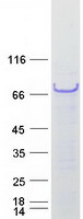
Figure 1. Western blot analysis of ASPH using anti-ASPH antibody (PB9478).
Electrophoresis was performed on a 5-20% SDS-PAGE gel at 70V (Stacking gel) / 90V (Resolving gel) for 2-3 hours. The sample well of each lane was loaded with 50ug of sample under reducing conditions.
Lane 1: Rat Brain Tissue Lysate,
Lane 2: Rat Liver Tissue Lysate,
Lane 3: HELA Whole Cell Lysate,
Lane 4: HEPG2 Whole Cell Lysate,
Lane 5: HEPA Whole Cell Lysate.
After Electrophoresis, proteins were transferred to a Nitrocellulose membrane at 150mA for 50-90 minutes. Blocked the membrane with 5% Non-fat Milk/ TBS for 1.5 hour at RT. The membrane was incubated with rabbit anti-ASPH antigen affinity purified polyclonal antibody (Catalog # PB9478) at 0.5 μg/mL overnight at 4°C, then washed with TBS-0.1%Tween 3 times with 5 minutes each and probed with a goat anti-rabbit IgG-HRP secondary antibody at a dilution of 1:10000 for 1.5 hour at RT. The signal is developed using an Enhanced Chemiluminescent detection (ECL) kit (Catalog # EK1002) with Tanon 5200 system. A specific band was detected for ASPH at approximately 100KD. The expected band size for ASPH is at 86KD.

Click image to see more details
Figure 2. IHC analysis of ASPH using anti-ASPH antibody (PB9478). ASPH was detected in paraffin-embedded section of Human Mammary Cancer Tissue. Heat mediated antigen retrieval was performed in citrate buffer (pH6, epitope retrieval solution) for 20 mins. The tissue section was blocked with 10% goat serum. The tissue section was then incubated with 1μg/ml rabbit anti-ASPH Antibody (PB9478) overnight at 4°C. Biotinylated goat anti-rabbit IgG was used as secondary antibody and incubated for 30 minutes at 37°C. The tissue section was developed using Strepavidin-Biotin-Complex (SABC)(Catalog # SA1022) with DAB as the chromogen.

Click image to see more details
Figure 3. IHC analysis of ASPH using anti-ASPH antibody (PB9478).
ASPH was detected in immunocytochemical section of A549 cell. Enzyme antigen retrieval was performed using IHC enzyme antigen retrieval reagent (AR0022) for 15 mins. The cells were blocked with 10% goat serum. And then incubated with 1μg/ml rabbit anti-ASPH Antibody ( PB9478) overnight at 4°C. Biotinylated goat anti-rabbit IgG was used as secondary antibody and incubated for 30 minutes at 37°C. The section was developed using Strepavidin-Biotin-Complex (SABC)(Catalog # SA1022) with DAB as the chromogen.

Click image to see more details
Figure 4. Flow Cytometry analysis of HeLa cells using anti-ASPH antibody (PB9478).
Overlay histogram showing HeLa cells stained with PB9478 (Blue line).The cells were blocked with 10% normal goat serum. And then incubated with rabbit anti-ASPH Antibody (PB9478,1μg/1x106 cells) for 30 min at 20°C. DyLight488 conjugated goat anti-rabbit IgG (BA1127, 5-10μg/1x106 cells) was used as secondary antibody for 30 minutes at 20°C. Isotype control antibody (Green line) was rabbit IgG (1μg/1x106) used under the same conditions. Unlabelled sample (Red line) was also used as a control.

Click image to see more details
Figure 5. Flow Cytometry analysis of U87 cells using anti-ASPH antibody (PB9478).
Overlay histogram showing U87 cells stained with PB9478 (Blue line). To facilitate intracellular staining, cells were fixed with 4% paraformaldehyde and permeabilized with permeabilization buffer. The cells were blocked with 10% normal goat serum. And then incubated with rabbit anti-ASPH Antibody (PB9478,1μg/1x106 cells) for 30 min at 20°C. DyLight488 conjugated goat anti-rabbit IgG (BA1127, 5-10μg/1x106 cells) was used as secondary antibody for 30 minutes at 20°C. Isotype control antibody (Green line) was rabbit IgG (1μg/1x106) used under the same conditions. Unlabelled sample without incubation with primary antibody and secondary antibody (Red line) was used as a blank control.
Protein Target Info & Infographic
Gene/Protein Information For ASPH (Source: Uniprot.org, NCBI)
Gene Name
ASPH
Full Name
Aspartyl/asparaginyl beta-hydroxylase
Weight
85863 MW
Superfamily
aspartyl/asparaginyl beta-hydroxylase family
Alternative Names
Aspartyl/asparaginyl beta-hydroxylase;1.14.11.16 ;Aspartate beta-hydroxylase;ASP beta-hydroxylase;Peptide-aspartate beta-dioxygenase;ASPH;BAH; ASPH AAH, BAH, CASQ2BP1, FDLAB, HAAH, JCTN, junctin aspartate beta-hydroxylase aspartyl/asparaginyl beta-hydroxylase|A beta H-J-J|ASP beta-hydroxylase|cardiac junctin|humbug|junctate|peptide-aspartate beta-dioxygenase
*If product is indicated to react with multiple species, protein info is based on the gene entry specified above in "Species".For more info on ASPH, check out the ASPH Infographic

We have 30,000+ of these available, one for each gene! Check them out.
In this infographic, you will see the following information for ASPH: database IDs, superfamily, protein function, synonyms, molecular weight, chromosomal locations, tissues of expression, subcellular locations, post-translational modifications, and related diseases, research areas & pathways. If you want to see more information included, or would like to contribute to it and be acknowledged, please contact [email protected].
Specific Publications For Anti-Aspartate beta hydroxylase/ASPH Antibody Picoband® (PB9478)
Hello CJ!
No publications found for PB9478
*Do you have publications using this product? Share with us and receive a reward. Ask us for more details.
Recommended Resources
Here are featured tools and databases that you might find useful.
- Boster's Pathways Library
- Protein Databases
- Bioscience Research Protocol Resources
- Data Processing & Analysis Software
- Photo Editing Software
- Scientific Literature Resources
- Research Paper Management Tools
- Molecular Biology Software
- Primer Design Tools
- Bioinformatics Tools
- Phylogenetic Tree Analysis
Customer Reviews
Have you used Anti-Aspartate beta hydroxylase/ASPH Antibody Picoband®?
Submit a review and receive an Amazon gift card.
- $30 for a review with an image
0 Reviews For Anti-Aspartate beta hydroxylase/ASPH Antibody Picoband®
Customer Q&As
Have a question?
Find answers in Q&As, reviews.
Can't find your answer?
Submit your question
7 Customer Q&As for Anti-Aspartate beta hydroxylase/ASPH Antibody Picoband®
Question
I was wanting to use your anti-Aspartate beta hydroxylase/ASPH antibody for ICC for mouse liver on frozen tissues, but I want to know if it has been validated for this particular application. Has this antibody been validated and is this antibody a good choice for mouse liver identification?
Verified Customer
Verified customer
Asked: 2020-02-10
Answer
It shows on the product datasheet, PB9478 anti-Aspartate beta hydroxylase/ASPH antibody has been tested for Flow Cytometry, IHC-P, ICC, WB on human, mouse, rat tissues. We have an innovator award program that if you test this antibody and show it works in mouse liver in IHC-frozen, you can get your next antibody for free.
Boster Scientific Support
Answered: 2020-02-10
Question
We are currently using anti-Aspartate beta hydroxylase/ASPH antibody PB9478 for human tissue, and we are content with the IHC-P results. The species of reactivity given in the datasheet says human, mouse, rat. Is it possible that the antibody can work on dog tissues as well?
Verified Customer
Verified customer
Asked: 2020-01-13
Answer
The anti-Aspartate beta hydroxylase/ASPH antibody (PB9478) has not been tested for cross reactivity specifically with dog tissues, though there is a good chance of cross reactivity. We have an innovator award program that if you test this antibody and show it works in dog you can get your next antibody for free. Please contact me if I can help you with anything.
Boster Scientific Support
Answered: 2020-01-13
Question
I am looking for to test anti-Aspartate beta hydroxylase/ASPH antibody PB9478 on mouse liver for research purposes, then I may be interested in using anti-Aspartate beta hydroxylase/ASPH antibody PB9478 for diagnostic purposes as well. Is the antibody suitable for diagnostic purposes?
Verified Customer
Verified customer
Asked: 2019-08-28
Answer
The products we sell, including anti-Aspartate beta hydroxylase/ASPH antibody PB9478, are only intended for research use. They would not be suitable for use in diagnostic work. If you have the means to develop a product into diagnostic use, and are interested in collaborating with us and develop our product into an IVD product, please contact us for more discussions.
Boster Scientific Support
Answered: 2019-08-28
Question
Will anti-Aspartate beta hydroxylase/ASPH antibody PB9478 work for ICC with liver?
O. Carter
Verified customer
Asked: 2017-06-19
Answer
According to the expression profile of liver, ASPH is highly expressed in liver. So, it is likely that anti-Aspartate beta hydroxylase/ASPH antibody PB9478 will work for ICC with liver.
Boster Scientific Support
Answered: 2017-06-19
Question
Does PB9478 anti-Aspartate beta hydroxylase/ASPH antibody work on parafin embedded sections? If so, which fixation method do you recommend we use (PFA, paraformaldehyde, other)?
R. Kulkarni
Verified customer
Asked: 2016-02-11
Answer
As indicated on the product datasheet, PB9478 anti-Aspartate beta hydroxylase/ASPH antibody as been tested on ICC. It is best to use PFA for fixation because it has better tissue penetration ability. PFA needs to be prepared fresh before use. Long term stored PFA turns into formalin, as the PFA molecules congregate and become formalin.
Boster Scientific Support
Answered: 2016-02-11
Question
See below the WB image, lot number and protocol we used for liver using anti-Aspartate beta hydroxylase/ASPH antibody PB9478. Please let me know if you require anything else.
F. Johnson
Verified customer
Asked: 2013-12-26
Answer
Thank you very much for the data. Our lab team are working to resolve this as quickly as possible, and we appreciate your patience and understanding! You have provided everything we needed. Please let me know if there is anything you need in the meantime.
Boster Scientific Support
Answered: 2013-12-26
Question
Is this PB9478 anti-Aspartate beta hydroxylase/ASPH antibody reactive to the isotypes of ASPH?
A. Jha
Verified customer
Asked: 2013-03-19
Answer
The immunogen of PB9478 anti-Aspartate beta hydroxylase/ASPH antibody is A synthetic peptide corresponding to a sequence at the C-terminus of human ASPH (726-758aa EVWQDASSFRLIFIVDVWHPELTPQQRRSLPAI), identical to the related mouse sequence. Could you tell me which isotype you are interested in so I can help see if the immunogen is part of this isotype?
Boster Scientific Support
Answered: 2013-03-19
