Click image to see more details
-

-
-
-
-
+1
Product Info Summary
| SKU: | PB10040 |
|---|---|
| Size: | 100 μg/vial |
| Reactive Species: | Human, Monkey |
| Host: | Rabbit |
| Application: | IF, IHC, ICC, WB |
Customers Who Bought This Also Bought


Product info
Product Name
Anti-ABP1/AOC1 Antibody Picoband®
SKU/Catalog Number
PB10040
PB1074 is an alternative SKU for this antibody, used in previous lots.
Size
100 μg/vial
Form
Lyophilized
Description
Boster Bio Anti-ABP1/AOC1 Antibody Picoband® catalog # PB10040. Tested in IF, IHC, ICC, WB applications. This antibody reacts with Human, Monkey. The brand Picoband indicates this is a premium antibody that guarantees superior quality, high affinity, and strong signals with minimal background in Western blot applications. Only our best-performing antibodies are designated as Picoband, ensuring unmatched performance.
Storage & Handling
Store at -20˚C for one year from date of receipt. After reconstitution, at 4˚C for one month. It can also be aliquotted and stored frozen at -20˚C for six months. Avoid repeated freeze-thaw cycles.
Cite This Product
Anti-ABP1/AOC1 Antibody Picoband® (Boster Biological Technology, Pleasanton CA, USA, Catalog # PB10040)
Host
Rabbit
Contents
Each vial contains 4 mg Trehalose, 0.9 mg NaCl and 0.2 mg Na2HPO4.
Clonality
Polyclonal
Isotype
Rabbit IgG
Immunogen
A synthetic peptide corresponding to a sequence at the N-terminus of human ABP1, different from the related mouse sequence by ten amino acids, and from the related rat sequence by eight amino acids.
*Blocking peptide can be purchased. Costs vary based on immunogen length. Contact us for pricing.
Cross-reactivity
No cross-reactivity with other proteins.
Reactive Species
PB10040 is reactive to AOC1 in Human, Monkey
Reconstitution
Add 0.2ml of distilled water will yield a concentration of 500ug/ml.
Observed Molecular Weight
85 kDa
Calculated molecular weight
85378 MW
Background of ABP1/AOC1
This gene encodes a metal-binding membrane glycoprotein that oxidatively deaminates putrescine, histamine, and related compounds. The encoded protein is inhibited by amiloride, a diuretic that acts by closing epithelial sodium ion channels. Alternatively spliced transcript variants encoding multiple isoforms have been observed for this gene. Catalyzes the degradation of compounds such as putrescine, histamine, spermine, and spermidine, substances involved in allergic and immune responses, cell proliferation, tissue differentiation, tumor formation, and possibly apoptosis. Placental DAO is thought to play a role in the regulation of the female reproductive function.
Antibody Validation
Boster validates all antibodies on WB, IHC, ICC, Immunofluorescence, and ELISA with known positive control and negative samples to ensure specificity and high affinity, including thorough antibody incubations.
Application & Images
Applications
PB10040 is guaranteed for IF, IHC, ICC, WB Boster Guarantee
Assay Dilutions Recommendation
The recommendations below provide a starting point for assay optimization. The actual working concentration varies and should be decided by the user.
Western blot, 0.1-0.5μg/ml, Human, Monkey
Immunohistochemistry(Paraffin-embedded Section), 2-5 μg/ml, Human
Flow Cytometry (Fixed), 1-3 μg/1x106 cells, Human
Positive Control
WB: human 293T whole cell, human HK-2 whole cell, monkey COS-7 whole cell
IHC: human colonic adenoma tissue, human placenta tissue, human renal cell carcinoma tissue
ICC/IF: T-47D cell
Validation Images & Assay Conditions
Click image to see more details
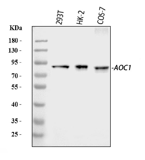
Figure 1. Western blot analysis of ABP1/AOC1 using anti-ABP1/AOC1 antibody (PB10040).
Electrophoresis was performed on a 5-20% SDS-PAGE gel at 70V (Stacking gel) / 90V (Resolving gel) for 2-3 hours. The sample well of each lane was loaded with 30 ug of sample under reducing conditions.
Lane 1: human 293T whole cell lysates,
Lane 2: human HK-2 whole cell lysates,
Lane 3: monkey COS-7 whole cell lysates.
After electrophoresis, proteins were transferred to a nitrocellulose membrane at 150 mA for 50-90 minutes. Blocked the membrane with 5% non-fat milk/TBS for 1.5 hour at RT. The membrane was incubated with rabbit anti-ABP1/AOC1 antigen affinity purified polyclonal antibody (Catalog # PB10040) at 0.5 μg/mL overnight at 4°C, then washed with TBS-0.1%Tween 3 times with 5 minutes each and probed with a goat anti-rabbit IgG-HRP secondary antibody at a dilution of 1:5000 for 1.5 hour at RT. The signal is developed using an Enhanced Chemiluminescent detection (ECL) kit (Catalog # EK1002) with Tanon 5200 system. A specific band was detected for ABP1/AOC1 at approximately 85 kDa. The expected band size for ABP1/AOC1 is at 85 kDa.

Click image to see more details
Figure 2. IHC analysis of ABP1/AOC1 using anti-ABP1/AOC1 antibody (PB10040).
ABP1/AOC1 was detected in a paraffin-embedded section of human colonic adenoma tissue. Heat mediated antigen retrieval was performed in EDTA buffer (pH 8.0, epitope retrieval solution). The tissue section was blocked with 10% goat serum. The tissue section was then incubated with 2 μg/ml rabbit anti-ABP1/AOC1 Antibody (PB10040) overnight at 4°C. Peroxidase Conjugated Goat Anti-rabbit IgG was used as secondary antibody and incubated for 30 minutes at 37°C. The tissue section was developed using HRP Conjugated Rabbit IgG Super Vision Assay Kit (Catalog # SV0002) with DAB as the chromogen.

Click image to see more details
Figure 3. IHC analysis of ABP1/AOC1 using anti-ABP1/AOC1 antibody (PB10040).
ABP1/AOC1 was detected in a paraffin-embedded section of human placenta tissue. Heat mediated antigen retrieval was performed in EDTA buffer (pH 8.0, epitope retrieval solution). The tissue section was blocked with 10% goat serum. The tissue section was then incubated with 2 μg/ml rabbit anti-ABP1/AOC1 Antibody (PB10040) overnight at 4°C. Peroxidase Conjugated Goat Anti-rabbit IgG was used as secondary antibody and incubated for 30 minutes at 37°C. The tissue section was developed using HRP Conjugated Rabbit IgG Super Vision Assay Kit (Catalog # SV0002) with DAB as the chromogen.

Click image to see more details
Figure 4. IHC analysis of ABP1/AOC1 using anti-ABP1/AOC1 antibody (PB10040).
ABP1/AOC1 was detected in a paraffin-embedded section of human renal cell carcinoma tissue. Heat mediated antigen retrieval was performed in EDTA buffer (pH 8.0, epitope retrieval solution). The tissue section was blocked with 10% goat serum. The tissue section was then incubated with 2 μg/ml rabbit anti-ABP1/AOC1 Antibody (PB10040) overnight at 4°C. Peroxidase Conjugated Goat Anti-rabbit IgG was used as secondary antibody and incubated for 30 minutes at 37°C. The tissue section was developed using HRP Conjugated Rabbit IgG Super Vision Assay Kit (Catalog # SV0002) with DAB as the chromogen.

Click image to see more details
Figure 5. IF analysis of ABP1/AOC1 using anti-ABP1/AOC1 antibody (PB10040).
ABP1/AOC1 was detected in an immunocytochemical section of T-47D cells. Enzyme antigen retrieval was performed using IHC enzyme antigen retrieval reagent (AR0022) for 15 mins. The cells were blocked with 10% goat serum. And then incubated with 5 μg/mL rabbit anti-ABP1/AOC1 Antibody (PB10040) overnight at 4°C. DyLight®488 Conjugated Goat Anti-Rabbit IgG (BA1127) was used as secondary antibody at 1:100 dilution and incubated for 30 minutes at 37°C. The section was counterstained with DAPI. Visualize using a fluorescence microscope and filter sets appropriate for the label used.
Protein Target Info & Infographic
Gene/Protein Information For AOC1 (Source: Uniprot.org, NCBI)
Gene Name
AOC1
Full Name
Amiloride-sensitive amine oxidase [copper-containing]
Weight
85378 MW
Superfamily
copper/topaquinone oxidase family
Alternative Names
Amiloride-sensitive amine oxidase [copper-containing];DAO;Diamine oxidase;1.4.3.22 ;Amiloride-binding protein 1;Amine oxidase copper domain-containing protein 1;Histaminase;Kidney amine oxidase;KAO;AOC1;ABP1, DAO1; AOC1 ABP, ABP1, DAO, DAO1, KAO amine oxidase copper containing 1 amiloride-sensitive amine oxidase [copper-containing]|amiloride binding protein 1 (amine oxidase (copper-containing))|amiloride-binding protein 1|amiloride-sensitive amine oxidase|amine oxidase copper domain-containing protein 1|diamine oxidase|histaminase|kidney amine oxidase
*If product is indicated to react with multiple species, protein info is based on the gene entry specified above in "Species".For more info on AOC1, check out the AOC1 Infographic

We have 30,000+ of these available, one for each gene! Check them out.
In this infographic, you will see the following information for AOC1: database IDs, superfamily, protein function, synonyms, molecular weight, chromosomal locations, tissues of expression, subcellular locations, post-translational modifications, and related diseases, research areas & pathways. If you want to see more information included, or would like to contribute to it and be acknowledged, please contact [email protected].
Specific Publications For Anti-ABP1/AOC1 Antibody Picoband® (PB10040)
Hello CJ!
No publications found for PB10040
*Do you have publications using this product? Share with us and receive a reward. Ask us for more details.
Recommended Resources
Here are featured tools and databases that you might find useful.
- Boster's Pathways Library
- Protein Databases
- Bioscience Research Protocol Resources
- Data Processing & Analysis Software
- Photo Editing Software
- Scientific Literature Resources
- Research Paper Management Tools
- Molecular Biology Software
- Primer Design Tools
- Bioinformatics Tools
- Phylogenetic Tree Analysis
Customer Reviews
Have you used Anti-ABP1/AOC1 Antibody Picoband®?
Submit a review and receive an Amazon gift card.
- $30 for a review with an image
0 Reviews For Anti-ABP1/AOC1 Antibody Picoband®
Customer Q&As
Have a question?
Find answers in Q&As, reviews.
Can't find your answer?
Submit your question
3 Customer Q&As for Anti-ABP1/AOC1 Antibody Picoband®
Question
Is this PB10040 anti-ABP1/AOC1 antibody reactive to the isotypes of AOC1?
Verified Customer
Verified customer
Asked: 2020-04-28
Answer
The immunogen of PB10040 anti-ABP1/AOC1 antibody is A synthetic peptide corresponding to a sequence at the N-terminus of human ABP1 (144-180aa STAEYALLYHTLQEATKPLHQFFLNTTGFSFQDCHDR), different from the related mouse sequence by ten amino acids, and from the related rat sequence by eight amino acids. Could you tell me which isotype you are interested in so I can help see if the immunogen is part of this isotype?
Boster Scientific Support
Answered: 2020-04-28
Question
We are currently using anti-ABP1/AOC1 antibody PB10040 for human tissue, and we are well pleased with the WB results. The species of reactivity given in the datasheet says human, rat. Is it possible that the antibody can work on monkey tissues as well?
Verified Customer
Verified customer
Asked: 2019-08-07
Answer
The anti-ABP1/AOC1 antibody (PB10040) has not been validated for cross reactivity specifically with monkey tissues, though there is a good chance of cross reactivity. We have an innovator award program that if you test this antibody and show it works in monkey you can get your next antibody for free. Please contact me if I can help you with anything.
Boster Scientific Support
Answered: 2019-08-07
Question
We appreciate helping with my inquiry over the phone. Here are the WB image, lot number and protocol we used for kidney using anti-ABP1/AOC1 antibody PB10040. Let me know if you need anything else.
Verified Customer
Verified customer
Asked: 2018-04-11
Answer
We appreciate the data. You have provided everything we needed. Our lab team are working to resolve your inquiry as quickly as possible, and we appreciate your patience and understanding! Please let me know if there is anything you need in the meantime.
Boster Scientific Support
Answered: 2018-04-11
