This website uses cookies to ensure you get the best experience on our website.
- Table of Contents
Counting cells before the staining procedure and the analysis/live sort is not up for debate! Cell numbers affect the staining quality, the FACS instrument reading, as well as the efficacy of any downstream assay, in case the cells are live sorted. Furthermore, cells are steadily lost during the staining procedure and the recovery of most sorters is a little above 50% − it is therefore imperative that enough cells are available at the start of the experiment to compensate for the inevitable losses. There are various methods available for counting cells:
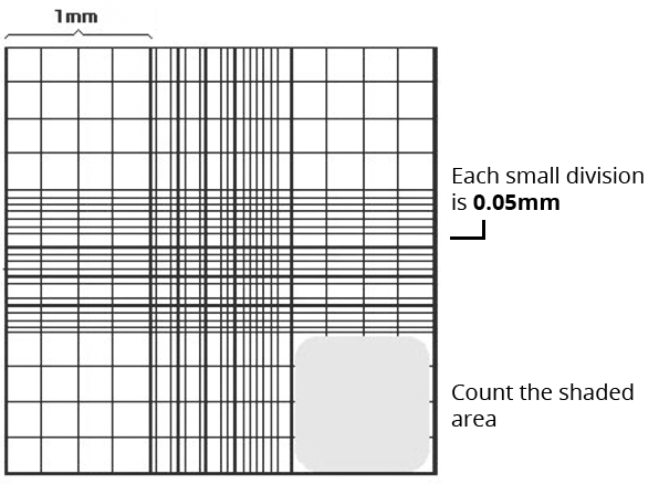
Hemocytometers are manual counting chambers and were first developed for blood cell counts. It consists of a thick glass slide onto which a gridded chamber is affixed. The four corner squares of the grid measure 1 mm x 1 mm and each square is further subdivided into 0.05 mm x 0.05 mm squares. A glass coverslip, made to specifications of the hemocytometer, can be placed 0.1 mm above the marked grid, thereby creating a space of known volume.
The cells are counted using a 4x or 10x objective lens over an area of 1 sq mm and the counting is repeated for a total of four such areas and the result is averaged. The formula for counting cells for each area is (average number of cells * 10 4 * dilution factor). To count the viable cells, use Trypan Blue exclusion method. Save some time and trouble with Boster’s ready-to-use Trypan Blue Assay Kit AR1175, see the kit below!.
Although the easiest method and least expensive, it is also labor intensive and allows for inaccuracies of the human eye. The image below depicts a typical hemocytometer counting grid.

They perform on the same principle as the hemocytometers but with the added advantages of speed and precision. Automated cell counter models have been developed by many different companies, so be sure to check their technical specifications. The only drawback of automated systems is that it cannot discern some irregular or unusually shaped cells as well as the human eye can.
These instruments do not measure the cells optically but through the electrical resistance. The principle is that cells have a greater resistance than the electrolyte solution in which they have been suspended; therefore, when a brief current is passed, the presence of a cell causes a brief change in resistance. It is this change that is detected by the machine and counted as a ‘cell’. The advantage of Coulter counters is the high speed, while a major drawback is the inability to distinguish between live and dead cells. Therefore its application is limited to performing counts of freshly isolated blood samples which hardly have any dead cells.
Be sure to weigh your options for the best results! For more flow cytometry technical resources, just click this button:
View FACS Resources*Note to educators: You are permitted to share Boster Bio's resources and PDFs on your class websites and lab websites. Please make sure to cite or link to the origin.
Here are some quick facts about bosterbio.com