This website uses cookies to ensure you get the best experience on our website.
- Table of Contents
Facts about Ubiquitin D.
Conjugation capacity triggered by UBA6. Promotes the expression of the proteasome subunit beta type-9 (PSMB9/LMP2).
Human | |
---|---|
Gene Name: | UBD |
Uniprot: | O15205 |
Entrez: | 10537 |
Belongs to: |
---|
No superfamily |
Diubiquitin; FAT10; FAT10diubiquitin; GABBR1; UBD; UBD-3; ubiquitin D; Ubiquitin-like protein FAT10
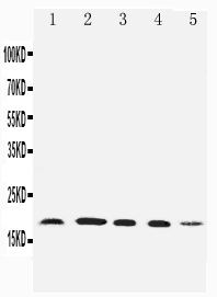
Mass (kDA):
18.473 kDA
Human | |
---|---|
Location: | 6p22.1 |
Sequence: | 6; NC_000006.12 (29555515..29559732, complement) |
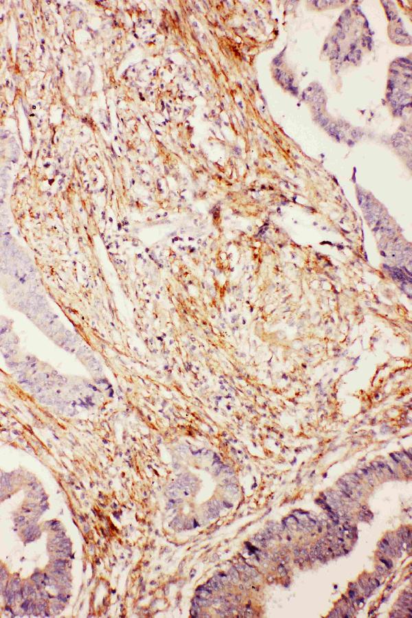
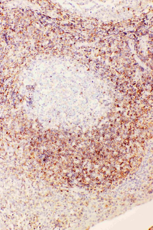
Constitutively expressed in mature dendritic cells and B-cells. Mostly expressed in the reticuloendothelial system (e.g. thymus, spleen), the gastrointestinal system, kidney, lung and prostate gland.
Nucleus. Cytoplasm. Accumulates in aggresomes under proteasome inhibition conditions.
One of the most popular ways to measure UBD markers is to amplify the mRNA or protein of interest. Boster bio offers high affinity primary antibodies that can be validated by Western Blotting. The UBD marker is a useful tool to detect and characterize the UBD-marker. Its high-affinity characteristics make it ideal in Western blotting experiments.
As a leading supplier of highly specific, specialized antibodies, Boster offers a variety of options to suit any research need. High-affinity primaries can be used in a variety if applications such as IHC and WB, FC, or picogram sensitivity ELISA kit kits. Each purchase of a primary antibody will also include a complementary one. BioVision Therapeutics high-affinity antibodies have been rigorously tested and are supported by an extensive technical support network.
In this study, tumor cell lines were inoculated on 24-well plates and treated for 20 minutes with 4% paraformaldehyde. After blocking the samples in 5% FBS for an hour, inoculating the primary antibodies was done. The inoculated primary antibodies were incubated at 4degC overnight. Bcl-2, B-STAT3, p-p65 and p-STAT3 were used to target. To determine the optimal antibody concentration, these secondary antibodies were incubated in the presence of a fluorescent-conjugated monoclonal antigen, 1:500. The samples of the antibodies were photographed at 554nm by a fluorescent microscope.
Monoclonal antibodies, as their names suggest, are single IgG proteins that bind to a single epitope, with high specificity and affinity. High-affinity antibodies, on the contrary, are more expensive and come with a low background. This makes them less common. High-affinity antibodies should be bought in large quantities to avoid waste of time.
A secondary antibody is often used with a primary antibody. This allows researchers different questions about a sample using different antibodies. A dual antibody can cross-react between the two so it's important that cross-reactivity is minimized. Alternately, you can use the primary antibody for both labeling and detection. This is a sensitive method that requires careful optimization to minimize cross-reactivity and maximize specificity.
Scientists place high importance on reproducibility of research antibody results. However, there are not universal standards. To ensure reproducible results, rigorous antibody validation should be carried out. Different immunoassays require different standards for antibody validation. This means that all antibodies must be validated for specificity and reproducibility according to the context in their use. Here are some guidelines for antibody validation
First, primary antibodies and secondaries should be validated in the same conditions. To identify cross-reactivity or potential problems with secondary antibody, a control blot should be used. Secondary antibodies should be distinguished from primary antibodies by their ability recognize different fluorophores or chemiluminescent signals. These guidelines are also outlined in the IWGAV recommendations. Boster’s primary antibodies are validated via Western blotting. It is an excellent method for evaluating the effectiveness.
Reproducibility of antibodies requires collaboration between users, vendors, and publishers. To determine the performance of an antibody, the user should design well-designed experiments. The vendor must supply high-quality antibodies as well as detailed disclosures about the validation procedures. The publisher can also establish guidelines for validation and enforce them. Together, these efforts will lead to improved reproducibility of antibodies. So, what can antibody researchers do?
Positive controls could include a lysate obtained from a cell culture expressing the target gene. If positive results are obtained, then the validation protocol was successful. Negative results can occur when the protein isn't expressed in the samples. Overexpression lysates serve as positive controls for routine tests, but are not used to evaluate antibody selectivity or offtarget binding.
Secondary antibodies may use fluorophore-conjugated primary antibodies to detect proteins. These antibodies have the fluorophores property to absorb light at one wavelength, and emit it at an alternate wavelength. With a single antibody, you can detect multiple proteins by using several fluorescent secondary antibodies. Primary antibodies are more sensitive and more specific than secondary antibodies.
The primary antibody anti-cofilin detects cofilin. It is a protein that is 19 kDa (or higher). The Odyssey blocking buffer reduces off-target binding further increasing its specificity. The same primary antibody can also be used in the determination of protein modifications. These antibodies are critical in the validation of Western Blotting.
UBD (Universal Biodiversity markers) is a reagent providing 68 different antibody types. The UBD marker can be used to determine the presence of an antibody in blood. UBD markers are available in many forms, including assays. They can also be used to identify specific proteins within the blood. In the following paragraphs we will explain how Boster’s UBDMarker works.
PMID: 9368598 by Bates E.E.M., et al. Identification and analysis of a novel member of the ubiquitin family expressed in dendritic cells and mature B cells.
PMID: 10200259 by Liu Y.-C., et al. A MHC-encoded ubiquitin-like protein (FAT10) binds noncovalently to the spindle assembly checkpoint protein MAD2.