This website uses cookies to ensure you get the best experience on our website.
- Table of Contents
Facts about Tumor necrosis factor receptor superfamily member 25.
Receptor for TNFSF12/APO3L/TWEAK.
Interacts directly with the adapter TRADD.Mediates activation of NF-kappa-B and causes apoptosis. May play a role in regulating lymphocyte homeostasis.
Human | |
---|---|
Gene Name: | TNFRSF25 |
Uniprot: | Q93038 |
Entrez: | 8718 |
Belongs to: |
---|
No superfamily |
APO3; Apo-3; Apoptosis-inducing receptor AIR; Apoptosis-mediating receptor DR3; Apoptosis-mediating receptor TRAMP; Death receptor 3; DR3; DR3member 12 (translocatingchain-association membrane protein); LARD; Lymphocyte-associated receptor of death; Protein WSL; Protein WSL-1; TNFRSF25; TRAMP; tumor necrosis factor receptor superfamily, member 25; WSL; WSL1; WSL-1
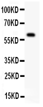
Mass (kDA):
45.385 kDA
Human | |
---|---|
Location: | 1p36.31 |
Sequence: | 1; NC_000001.11 (6460786..6466195, complement) |
Abundantly expressed in thymocytes and lymphocytes. Detected in lymphocyte-rich tissues such as thymus, colon, intestine, and spleen. Also found in the prostate.
[Isoform 1]: Cell membrane; Single-pass type I membrane protein.; [Isoform 2]: Cell membrane; Single-pass type I membrane protein.; [Isoform 9]: Cell membrane; Single-pass type I membrane protein.; [Isoform 11]: Cell membrane; Single-pass type I membrane protein.; [Isoform 3]: Secreted.; [Isoform 4]: Secreted.; [Isoform 5]: Secreted.; [Isoform 6]: Secreted.; [Isoform 7]: Secreted.; [Isoform 8]: Secreted.; [Isoform 10]: Secreted.; [Isoform 12]: Secreted.
Boster Bio is the best TNFRSF25 marker. This primary antibody is high-affinity and can recognize the AMPK–mTOR–Ulk1/2 AMPK-mTOR-Ulk1/2, which regulates cellular autophagy. Its unique design enables you to detect autophagy from a wide variety samples, including tissues and organs.
These antibodies can have many forms. They are affinity-based, high-affinity antibodies that recognize the TNFRSF25 Marker. The Fc domain peptides contain two polypeptides: one heavy, one light. The disulfide bond joins the two chains. Additionally, antibodies have a higher affinity for neutral pH than antibodies that are designed to recognize acidic.
Some antibodies are composed of multiple polypeptides capable of binding the target protein. Sometimes, the host cell has expression vectors that allow the production of the antibody. These polypeptides are either from the immunoglobulin heavier chain or the lighter chain. These polypeptides may have a mutant or variant Fc domain, and may bind to FcRn with enhanced affinity at acidic pH.
Some compositions may also contain engineered Fcdomains. These polypeptides are mutant versions Fc domains of wild-type human IgG1 antibodies. These antibodies not only have enhanced affinity but also have increased serum half lives in transgenic mice. They've been tested in primates and humans. The antibodies bearing the "YTE” variant are currently being evaluated at several clinical trials.
The AMPK-mTOR-ULK1/2 axis controls autophagy and mTORC1 negatively regulates its activity. Both AMPK1 and mTORC1 phosphorylate ULK1 while both regulate ULK1. For proper functioning, the interaction between these two determinants of Autophagy is crucial. An abnormal accumulation of autophagy-related protein results when both ULK1 and AMPK are not present in cells.
The AMPK mTOR ULK1/2 axis of eukaryotic catabolic pathways allows for the collection cellular waste. This contributes towards homoeostasis. These pathways are implicated in several human diseases, including cancer and neurodegeneration. They may also be promising therapeutic targets. The following sections detail the roles of each of these axes.
Autophagy is controlled by AMPK-mTOR/ULK1/2 by phosphorylating ULK1. The kinase links energy sensing and mitophagy. AMPK phosphorylates ULK1 at Ser-317 and Ser-555, while ULK1 does not at Ser-7777. The AMPK mTOR ULK1/2 regulates ULK1 by preventing degradation of cells and components.
In a recent study, we reported that phosphorylation of ULK1 at Ser757 is critical for autophagy. Atherosclerosis can be prevented by activating AMPK. It is a critical step in normal cardiovascular function. Activating AMPK stops the development of atherosclerosis. It also promotes the efferocytosis and cholesterol.
By inhibiting mTOR, ULK1 and macroautophagy are activated. In addition, inhibition of LRRK2 kinase induces autophagy in cells by phosphorylating ULK1 on Ser758. LRRK2 inhibition leads to mTOR-ULK1/2 pathway inhibition. In addition, prolonged inhibition of LRRK2 causes activation a feedback loop that suppresses ULK1 or mTOR.
Activation mTOR–ULK1 under LEP and EE results with the restoration ULK1 phosphorylation. Torin-1 is able to inhibit ULK1 phosphorylation in H4 cells but not LRRK2 inhibition. LRRK2 inhibition was similar to LRRK1 inhibitors in terms of its effects on autophagy. However LRRK2in1 did not show any mTOR/ULK1/ULK1 interaction during cell culture.
The initiation of mitophagy and its progression after mitochondrial injury is controlled by the AMPK/ULK1 axis. AMPK activation at ULK1 via PINK1-dependent phosphorylation Parkin required for rapid maximal Parkin phosphorylation with PINK1. Although AMPK was previously thought not to be involved in mitophagy at all, previous studies have not excluded any other targets/functions for AMPK during the initial hour.
Autophagy starts with the AMPK mTOR-ULK1 axis. It controls the synthesis ubiquitin dependent proteins and the activation dopaminergic neural cells. AMPK-mTOR-ULK1/2 axis also controls innate immune responses. Further research is needed regarding the role played by ULK1 in activating Parkin.
LEP saved myocardial function during exercise-induced myocardial infarction and reduced myocardial damage. LEP decreased the effect of autophagy inhibitions in the W+LEP+EE-group. LEP-treated cells also showed protection from myocardial damage, and HBFP-positive staining revealed a crimson-red patchy appearance.
Boster Bio TNFRSF25 identifies a cell’s capacity to enter autophagy. This is a highly efficient method to recycle domestic cellular components for nutrients and energy. Autophagy is crucial for cell homeostasis, stress responses, as well as multiple proteins that play key roles in bone cell survival and osteoblast growth.
Researchers who are interested using this marker to aid their research are encouraged not only to submit results but also samples, species, and applications. Boster Bio will provide links to their published resources. These resources are available to researchers from any country or institution. These resources may be shared with fellow researchers or students at no cost if they cite Boster Bio and link to it. Researchers from all over the world can use Boster Bio's TNFRSF25 marker in order to analyze autophagy.
PMID: 8934525 by Kitson J., et al. A death-domain-containing receptor that mediates apoptosis.
PMID: 8875942 by Chinnaiyan A.M., et al. Signal transduction by DR3, a death domain-containing receptor related to TNFR-1 and CD95.