This website uses cookies to ensure you get the best experience on our website.
- Table of Contents
30 Citations 5 Q&As
12 Citations 16 Q&As
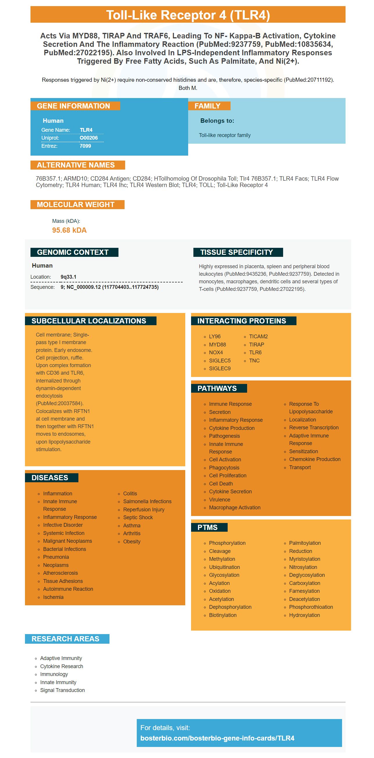
Facts about Toll-like receptor 4.
Responses triggered by Ni(2+) require non-conserved histidines and are, therefore, species-specific (PubMed:20711192). Both M.
Human | |
---|---|
Gene Name: | TLR4 |
Uniprot: | O00206 |
Entrez: | 7099 |
Belongs to: |
---|
Toll-like receptor family |
76B357.1; ARMD10; CD284 antigen; CD284; hTollhomolog of Drosophila toll; tlr4 76B357.1; TLR4 facs; TLR4 flow cytometry; TLR4 human; TLR4 ihc; TLR4 western blot; TLR4; TOLL; toll-like receptor 4
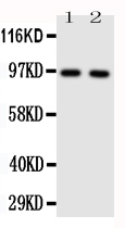
Mass (kDA):
95.68 kDA
Human | |
---|---|
Location: | 9q33.1 |
Sequence: | 9; NC_000009.12 (117704403..117724735) |
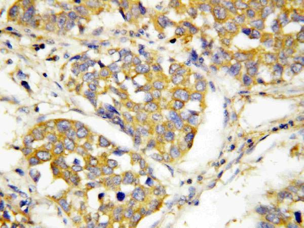
Highly expressed in placenta, spleen and peripheral blood leukocytes (PubMed:9435236, PubMed:9237759). Detected in monocytes, macrophages, dendritic cells and several types of T-cells (PubMed:9237759, PubMed:27022195).
Cell membrane; Single-pass type I membrane protein. Early endosome. Cell projection, ruffle. Upon complex formation with CD36 and TLR6, internalized through dynamin-dependent endocytosis (PubMed:20037584). Colocalizes with RFTN1 at cell membrane and then together with RFTN1 moves to endosomes, upon lipopolysaccharide stimulation.
TLR4 detection and AAD detection could be biomarkers used to detect Alzheimer's. What are these biomarkers and how can you use them for research? These questions will be explored in this article. We'll also discuss the importance and role of TLR4 as well as its agonist, PE6. If you are a researcher looking to find TLR4 biomarkers, this is the place for you.
Molecular Dynamics simulations have been used successfully to predict the docking energy for oleuropein with target protein. In these simulations, the energy was measured at 250,000 cal/mol and 100 times inside the binding site. The AutoDockTools software was used to analyze the docking data. Using Biovia Discovery Studio Visualizer, the best scoring pose was selected for further analysis.
CD14 can affect the TLR4 response of LPS. Some Gram-negative bacteria produce RLPS, which lacks an O–polysaccharide chains and bears incomplete oligosaccharides. Most E.coli strains (including the K235 strain) produce sugar-linked smooth LPS. LPS-induced signaling was activated in mice that had a premature stop codon at their Cd14 genes.
The solid phase of SP-A bound MD-2 in solid phase and TLR4 by SP-A bound MD-2. They had a similar affinity to TLR4, but different KD values. The SP-A-TLR4 interactions required CRD, oligomerization, and neck domain. These interactions were further explored using an anti-sTLR4 antibody (mAb–sTLR4). After performing this analysis, the SP-A-MD-2 complexes were immunoprecipitated using an anti-sTLR4 antibody.
After 20 ns, TLR4/MD-2 is stable. The ligand structure and the protein structure are stable up to 50 ns, with a slight deviation after 60. These results are similar with those seen for other complexes. These results indicate that the TLR4/MD-2 compound is stable for the longest duration of simulation. It may take longer simulations before the binding energies of the protein/ligand are regulated.
TLR4 is a transmembrane glycoprotein of type I, 100 kD that belongs to the Toll-Like Receptor Family. The receptor interacts directly with LPS by binding to its membrane and activating two distinct signaling pathways. These responses are controlled by the LPS-ligand combination. Monomeric receptors are favoured by antagonistic LPS. Monomeric LPS will be a monomeric receptor.
After NPM1 was added to cultures of Blue hTLR4 cells, the docking experiments were repeated three times. Quanti-Blue reagent is used to measure SEAP activity at 655 nm. This allows us to monitor the interaction of TLR4 with NPM1. The experiments were replicated three times and results were consistent. A decamer between NPM1 and TLR4 was detected.
In neutropenic patients, TLR4 may play a role in regulating inflammatory reactions. It is also implicated in the regulation of stem cell niches and immune reactivity in allogeneic stem cell transplant recipients. It also regulates bone marrow stromal cell neighbors and is associated with angiogenesis. TLR4 signals several physiological functions that are essential for normal immune function.
Both molecular dynamics simulations and molecular Docking showed that the two proteins had a stable interaction. To simulate the interaction between these molecules, molecular docking was used. The design construct has a high affinity towards TLR4 but is highly immunogenic. More research is required. The proposed TLR4/MD-2 anti-ants may be effective in treating various diseases.
According to the authors, serum MMP9 levels and TLR4 levels were related to ECOG performance status. Statistically significant differences were found between patients in ECOG 2 and ECOG 0. These results did not show any association with AAD histological type or treatment response. Future studies should explore the contribution of these proteins in biomarkers that help identify AAD.
MMPs and TLR4 both have been shown to inhibit elastase, as well as other proteins. Patients with AAD are more likely than others to have TLR4. MMPs are present in the blood but other proteins could inhibit them. McCawley and Matrisian suggest that these non-ECM proteins could be biomarkers to identify AAD.
The researchers also found that TLR4 as well as serum MMP9 levels were associated to AAD risk in Han people. This finding could have implications for understanding TLR4's role in the pathophysiology and treatment of AAD. TLR4 is an important pattern-recognition and initiation protein in the signal transduction pathway. TLR4-mediated Signaling is also important in maintaining aortic Homeostasis. A disease of the aorta is AAD.
Recent structural analyses have led the development of synthetic matrixin inhibitors. However, clinical trials have been inconclusive. It is possible that the inhibitors may not be effective in blocking the target enzymes. There are 23 MMPs in humans. The clinical trials on advanced patients may not be accurate. Future challenges include the development and testing of inhibitors for each specific MMP.
IPA also showed increased inflammation and expressions of certain serum proteins like suPAR, von Willebrand factors, tPA and suPAR. These changes were also related to the severity of the disease. In addition, SOMAscan intensity was significantly higher in patients who survived or died. These levels were higher in patients who survived than in those who died.
TLR4 or DC-SIGN could also be biomarkers for APD detection. They are two protein groups that are expressed by alveolar mammalian macrophages. They are useful biomarkers because they are associated to increased phagocytosis. TLR4 (serum MMP9) and serum MMP9 are potential biomarkers for diagnosing AAD.
Several recent reports on the effects of hypoxia and oxidative stress on neuronal cell death are presented in this review. The review will include discussion of biomarkers, clinical implications, and future perspectives. They could be helpful in developing clinical trials. The article also discusses recent advancements in epilepsy biomarkers.
The study also found that patients suffering from COVID-19 had higher rates of ARDS when they had high MMP9 levels and TLR4 levels. Patients with type-2 diabetes may not express enough macrophage proteins. This may make them more vulnerable to infection and disease severity. In addition, these two biomarkers are associated with increased glucose variability.
To identify TLR4 receptors in human cells, we used the Detection of TLR4 ligand PE6 as the target. This TLR4 ligand interacts to the surface immune receptor TLR4 in order to stimulate a strong secretion proinflammatory cytokines. We have shown that PE6 can induce the secretion of proinflammatory substances in DTLR4 knockout cells and DTLR2-knockout.
First, we performed protein-protein docking between the PE6 and TLR4 proteins using a docking algorithm. Next, we balanced electrostatic, hydrophobic and balanced scores to find the best model. This model was then sent to PDBPisa for analysis of protein-protein interactions. PE6 was found strongly to induce apoptosis, inhibit the autophagy process, and activate caspase-3.
Scientists have faced difficulties in detecting PE6 using thermal ramping. As a control, we used non-radioactive (dsDNA) to evaluate the stability. To verify that the TLR4-ligand compounds were viable, we used LPS (500ng/m) as a negative control. Before each reading, we pre-incubated PE6 (4 mM with 10 mMDNA).
PE6 inhibits autophagy of RAW264.7-cells. PE6 also inhibits formation of punctate foci associated with LC3BII. These results confirm previous experiments which showed that PE6 has an inhibitory effect. PE6 inhibits the initiation and progression of autophagy via inhibition of MtorC1 or suppression of ULK1.
TLR4 ligands can be detected by mAbs, which is an essential analytical tool in many fields of biology. TLR is one of a number of receptors that activates the immune system and detects pathogens. TLR activation in excess has been linked with several inflammatory and infectious diseases. PE6 also inhibits autophagy by repressing the autophagy initiating protein ULK1. Molecular studies show that TLR9's C terminal fragment is functional.
TLR4 model and PE6 model were created by ClusPro protein–protein docking. The colored bar indicates the coloration of PE6 from its N-terminal to C-terminal domain. Toll-like receptors play an essential role in the initiation and progression of inflammatory diseases. Toll-like receptor antagonists are useful in curing infectious diseases and preventing the development of inflammation.
Recent research has shown that PE6 increases expression ER stress markers such as Chop and Calnexin and promotes the production pro-apoptotic substances. This causes macrophages to die and facilitates mycobacterial cargo spreading. In addition to enhancing the inflammatory response, PE6 modulates the proinflammatory signaling cascade and cell death pathways.
After being incubated with PE6, macrophages were exposed for 30 minutes to M.TB. In the presence of PE6 (a TLR4 agonist). Afterwards, protein levels were determined using immunofluorescence microscopy. Also, P50 as well as P65 were found in the cells following treatment with PE6. These results suggest that PE6 activates P65. This is only one aspect, however, of how PE6 acts on macrophages.
PMID: 9237759 by Medzhitov R., et al. A human homologue of the Drosophila Toll protein signals activation of adaptive immunity.
PMID: 9435236 by Rock F.L., et al. A family of human receptors structurally related to Drosophila Toll.
Tetramethylpyrazine attenuates endotoxin-induced retinal inflammatio...
*Showing only the more recent 20. More publications can be found for each product on its corresponding product page