This website uses cookies to ensure you get the best experience on our website.
- Table of Contents
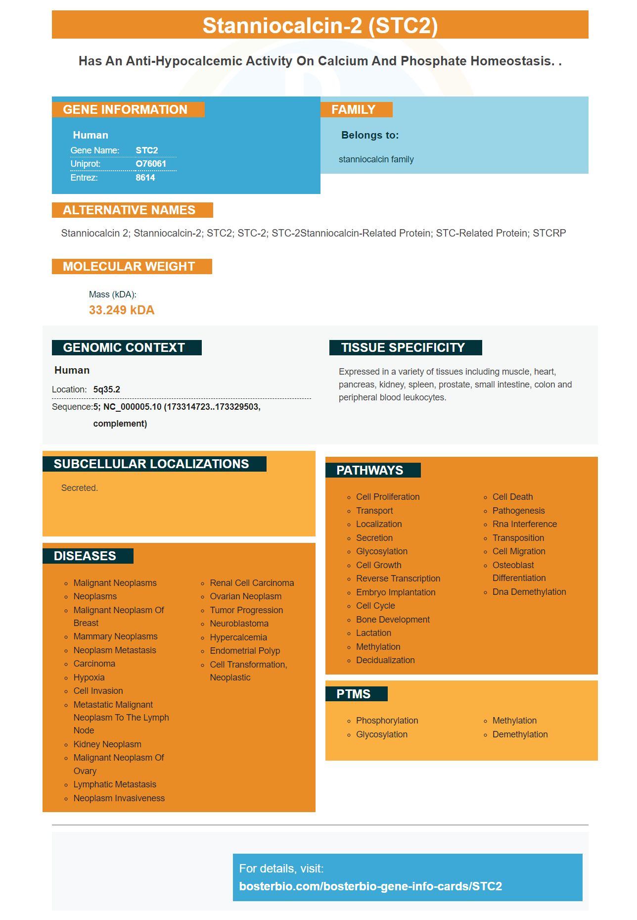
Facts about Stanniocalcin-2.
Human | |
---|---|
Gene Name: | STC2 |
Uniprot: | O76061 |
Entrez: | 8614 |
Belongs to: |
---|
stanniocalcin family |
Stanniocalcin 2; stanniocalcin-2; STC2; STC-2; STC-2Stanniocalcin-related protein; STC-related protein; STCRP
Mass (kDA):
33.249 kDA
Human | |
---|---|
Location: | 5q35.2 |
Sequence: | 5; NC_000005.10 (173314723..173329503, complement) |
Expressed in a variety of tissues including muscle, heart, pancreas, kidney, spleen, prostate, small intestine, colon and peripheral blood leukocytes.
Secreted.
This article will show you how to optimize your experiments by using the Boster bio: Best Uses Of MUC2C System. We'll discuss how STC2 regulates a series of biological processes, including cell proliferation and apoptosis. This article will also cover how miR-184 regulates this gene, as well as other questions that you may have. No matter what your experiment design is, there are some tips or guidelines that you should be aware of.
STC2 is part of the stanniocalcin famiy. It regulates many biological processes, including cell proliferation and insulin-like factor (IGF). This protein prevents apoptosis which is essential for cell survival. It also inhibits oxidative and pro-survival stress. In vitro, STC2 regulates the differentiation of human dental pulp cells.
STC2 contributes to the resolution of acute lung injury (ALI), but its role in the resolving of the condition is unclear. Recent studies have shown that STC2 promotes MSC formation, which is a cell type that promotes tissue repair and inflammation resolution. STC2 may also be involved in immunomodulatory and anti-apoptotic functions. However, more research is needed to establish a causal role of STC2.
STC2 regulates a variety of physiological processes. STC2 has also been shown in human nucleus pilutosus cells to influence apoptosis. This study also showed that STC2 affects miR184 which is a gene responsible for controlling cell proliferation. SiRNAs that target miR184 can induce apoptosis in cancer cells.
The CYP4F2 gene, which controls the proliferation and migration of liver cancer cells, has also been implicated in HCC tumorigenesis. The research team concluded that this gene is an important biomarker and could have prognostic value. This study was supported by the China Postdoctoral Science Fund and the National Natural Science Young Foundation. This makes it extremely important to understand the role of STC2 for HCC treatment.
MiR-184 targets STC2, an important gene involved in cell cycle control. The inhibition of STC2 results in increased apoptosis, and increased chemosensitivity. MiR-184 also inhibits the activity a protein called LINGO1. MiR-184 is a new small RNA that regulates LINGO1's function.
The glucose level in the blood is critical for STC2's cellular function. ER stress results in impaired glucose uptake and impairs ATP manufacturing. Low glucose also causes protein glycosylation to decrease, leading to misfolded proteins. Further research is needed to understand the protein's functions. However, the results support the idea that STC2 may be a general stress-responsive factor.
STC2 overexpression is positively associated with tumour growth, invasion and metastasis as well as patient prognosis. Its potential as both a biomarker for treatment and therapeutic target is also highlighted by the study. This review discusses STC2, its biological functions and future perspectives.
In addition to its effects on cell proliferation and migration, miR-184 also inhibits invasion and migration of RB cells. It also inhibits apoptosis. miR-184 mimic inhibits the expression of SLC7A5 by decreasing the ratio of EdU-positive cells in RB cells. miR-184 also inhibits STC2, an essential protein for myelination.
You can determine if the STC2 marker is responsible for cancer cell proliferation and survival by using human cell lines. STC2 activates the PI3K/AKT/Snail signaling pathway. This marker promotes proliferation and passage through G1/S cells. Furthermore, it inhibits apoptosis in carcinoma cells. STC2 not only promotes proliferation but also regulates gene expression of BCL2.
Ribobio Company has developed siRNA-STC2 which suppresses STC2 expression, and inhibits its activity, in order to better understand the regulatory role played by STC2 in EECs. An inhibitor of miR184 also reduced the effects of STC2 by decreasing miR-184 expression. The combination of STC2 + miR-184 promoted EEC proliferation and apoptosis.
The vital role of activating the RAS/RAF/MEK/ERK pathways in cell proliferation and apoptosis is played by EECs. STC2 plays an important role in cancer cell proliferation and also controls apoptosis within colorectal cells. MiR-184, on the other hand, inhibits STC2 but activates apoptosis-inducing factors p53 and p53. These results suggest that STC2 plays an essential role in regulating colorectal carcinoma cell survival.
MiR-184 regulates gene expression of the STC2 target gene. It is found in 293T cells where STC2 and miR-184 are co-transfected. After transfection, the cell activity was measured using double luciferase assay. Furthermore, miR-184 reduced STC2 expression by dairy goat EECs. It is important that miR184 inhibits RAS/RAF/MEK/ERK Signaling Pathway.
Researchers found that Hep3B cells overexpressing the STC2 marker caused apoptosis in a recent study. They used an Annexin V-allophycocyanin apoptosis detection tool from Merck KGaA or Sigma-Aldrich. Cells treated by CYP4F2 with an empty vector control were less likely to undergo apoptosis compared to cells treated with the positive control. This supports the hypothesis of CYP4F2 acting as a crucial regulator of Hep3B cell apoptosis.
To study the effects of the STC2 marker upon tumor growth, the research team used a nude mouse xenograft model to study the tumor. The STC2 marker targets protein-kinase B, and extracellular signal-regulated Kinase. It was therefore chosen as a potential DEG. The same goes for miR-190 which is an inhibitor in proliferation, angiogenesis and EMT.
HNSCC cell metastasis is promoted by activation of the STC2 signaling pathway. This signaling pathway plays vital role in the survival of cancer-cells. We examined the role STC2 played in tumor angiogenesis. Cell migration and invasion were also investigated. We also found that STC2 is involved in the regulation of survival of HNSCC cells.
STC2 inhibits cell apoptosis. Western blotting confirmed the presence of STC2 on HNSCC cell line cells. To determine the effects of STC2 in HNSCC cells' proliferation, we used the Cell Counting Kit-8 reagent. Our data shows that STC2-transfected cells show enhanced colony formation.
STC2 may regulate Snail. This is an essential protein in tumorigenesis. STC2 regulates Snail via AKT/PI3K signaling and promotes the invasion and migration of gliomas cells. This suggests that Snail could be a therapeutic target for glioma. The safety of a therapeutic drug targeting Snail is also demonstrated by the study.
The study found that the STC2 gene was induced by HIF1a, and modulated by two cofactors: histone acetyltransferase 300 and histone deacetylase 7 The STC2-coding DNA was placed into a lentivirus virus vector. These lentiviruses could be generated from 293T packing cells and infected target line cell lines. Cal-27, Tca-8113 were the cells containing STC2 cDNA.
Ligand-membrane receptor (ligand-membrane) binding activates the PI3K/AKT/snail signalsing pathway. In addition, a subunit called GRB2 binds the SOS protein. In turn, SOS activates the RAS and activates the p110a subunit. STC2 activation can also be caused by phosphorylation (Akt) of the signaling pathway.
PMID: 9723890 by Chang A.C.-M., et al. Identification of a second stanniocalcin cDNA in mouse and human: stanniocalcin 2.
PMID: 9753616 by Ishiabshi K., et al. Molecular cloning of a second human stanniocalcin homologue (STC2).