This website uses cookies to ensure you get the best experience on our website.
- Table of Contents
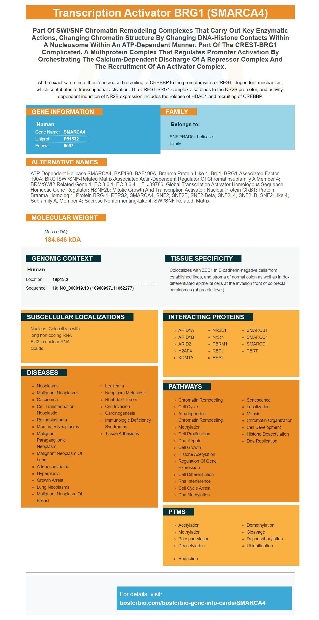
Facts about Transcription activator BRG1.
At the exact same time, there's increased recruiting of CREBBP to the promoter with a CREST- dependent mechanism, which contributes to transcriptional activation. The CREST-BRG1 complex also binds to the NR2B promoter, and activity-dependent induction of NR2B expression includes the release of HDAC1 and recruiting of CREBBP.
Human | |
---|---|
Gene Name: | SMARCA4 |
Uniprot: | P51532 |
Entrez: | 6597 |
Belongs to: |
---|
SNF2/RAD54 helicase family |
ATP-dependent helicase SMARCA4; BAF190; BAF190A; brahma protein-like 1; Brg1; BRG1-associated factor 190A; BRG1SWI/SNF-related matrix-associated actin-dependent regulator of chromatinsubfamily A member 4; BRM/SWI2-related gene 1; EC 3.6.1; EC 3.6.4.-; FLJ39786; global transcription activator homologous sequence; homeotic gene regulator; hSNF2b; Mitotic growth and transcription activator; nuclear protein GRB1; Protein brahma homolog 1; Protein BRG-1; RTPS2; SMARCA4; SNF2; SNF2B; SNF2-beta; SNF2L4; SNF2LB; SNF2-like 4; subfamily a, member 4; sucrose nonfermenting-like 4; SWI/SNF related, matrix
Mass (kDA):
184.646 kDA
Human | |
---|---|
Location: | 19p13.2 |
Sequence: | 19; NC_000019.10 (10960997..11062277) |
Colocalizes with ZEB1 in E-cadherin-negative cells from established lines, and stroma of normal colon as well as in de-differentiated epithelial cells at the invasion front of colorectal carcinomas (at protein level).
Nucleus. Colocalizes with long non-coding RNA Evf2 in nuclear RNA clouds.
The SMARCA4 marker is a versatile gene expression biomarker used to identify mutations in various human genes. We will explore its most important uses in this article. We will also look at primers that were employed to identify this gene, including ChIP–Seq results from MDA-MB-231 cell lines. We will also address small molecule with direct-acting properties (DASMs) that target their bromodomains.
This kit includes two specific RT-PCR primers, each designed for one of these genes. The forward primer is a part of the EcoRI recognition site, as well as the ATG codeon for initiation. The coding sequence for human MEIS1 was discovered using the NCBI RefSeq Nm_002398 reference genome. Codons 2-6 form the code sequence. The reverse primer includes an TAG stopcodon as well as codons 385-390.
HOXA9 and MEIS1 are genes that regulate cell growth and differentiation in skeletal muscles. In denervated muscles, satellite cells do not develop into myotubes. However, their expression is significantly higher than that of healthy muscles. This observation was confirmed by the research conducted by Maier and Bornemann (2003) and is consistent with ours. These studies demonstrate that HOXA9 is responsible for regulating the fate of denervated muscle cells as well as the direction of satellite cell development.
miR-182 targets the 3'UTR for the HOXA9 3'UTR. This target sequence was constructed using pMIR–REPORT vector, which is located between SpeI sites and HindIII. The mutant site was designed with 5'-GCTCTAGCCTTGAGGCTTAT-3' primers. Both the mutant and wt constructs were transfected into HeK-293T cells.
Potentially cancer-related genes have been discovered in MEIS1 and HOXA9 transcription factors. HOXA10 regulates embryonic uterine growth and the receptivity of adult endometrial endometrial. TALE (three-amino acid loop extension) family homeobox genes increase the activity of target genes and enhance their specificity. Additionally TALE proteins act as cofactors of HOX.
ChIP-Seq analysis in MDA-MB-231 cancer cells provides a powerful genomic assay. This method permits the identification of key genes that play a role in the development of breast cancer. Two cell lines were utilized in the study: MDAMB-231 cells and the LM2-4175 cell. The chromatin immunoprecipitated out of these cells was sonicated , then separated into 200-500 bp fragments. The data were then analyzed by peak calling. The peak was defined as the region that had q0.05. The inputs were also used as control data. Each peak was assigned the nearest RefSeq gene.
The study also revealed a wide expression profile for breast cancer cells. Multiple histone modifications affected the expression of genes in a coordinated manner. ChIP-Seq results revealed specific cis-elements or TFs that are involved in lung metastasis. In addition to lung metastasis the study also revealed the transcriptional profile of a subset of breast cancer genes, which include HER2, MCF-7 and HER2.
The results indicated that JMJD6 and EZH2 regulate the same gene cause. Both proteins are chromatin modifiers but their profiles differ. Both JMJD6 and EZH2 have overlapping peaks within their profiles, but their peaks are not close to 100 kb from each other. The peaks of these two proteins coincided around the same binding sites.
The enrichment plots of MCF7 cells and MDAMB-231 cells revealed different patterns in the distribution of the regions that are expressed. The high-frequency peaks of MCF7 were larger and more prominent than those of MDA-MB-231 cells. In contrast, the peaks for MCF7 and MDAMB-231 cells were significantly more enriched with cancerous samples.
Two MDA-MB-231 knockdown knockdown clones stable were used to collect gene expression data, and two control clones were also used. The data were processed using RMA. This was then implemented within the Bioconductor program. The resulting PPI networks then had to be examined. The analysis of the gene expression revealed critical genes involved in breast cancer. In this study, two genes from MZF1 were identified as involved in cancer progression.
The results of this study show that ChIP-Seq enrichment of the transcription start site (TSS) was higher in LM2-4175 cells than MDA-MB-231 cells. This is in part due to the increase in the HMT SETD7 and the downregulation of KDM2A. H3K27ac enrichment in LM2-4175 cells was lower however.
Gene expression FCs were highly related to changes in the state of the promoter. Genes whose promoters changed from active to repressive states had the most average FC and those with transitions from active to none states were lower. In contrast, genes with high FCs showed a higher likelihood to metastasize. Additionally, the gene expression of ZBTB7A has been associated with the risk of breast cancer.
The quality of the localization data provided by ChIP-Seq analysis was high. High mapping efficiency and a low PCR duplication rate, and good reproducibility between biological replicates are indicators of high quality localization. These characteristics were crucial to identifying lung-related regions of metastasis. Although it is important in lung development, however the majority of studies haven't been able to establish the role played by LMO4 in lung metastasis.
Using a strategy known as direct-acting small molecule targeting their bromodomains, CellCentric discovered an agent that inhibits the p300/CBP protein through its conserved bromodomain pockets. While other pockets of the protein could be targeted as well as binding to bromodomains can provide the balance between selectiveness and potency. CellCentric has now identified Inobrodib, the first inhibitor of p300/CBP, as well as a series of possible future drugs.
PMID: 8232556 by Khavari P.A., et al. BRG1 contains a conserved domain of the SWI2/SNF2 family necessary for normal mitotic growth and transcription.
PMID: 8208605 by Chiba H., et al. Two human homologues of Saccharomyces cerevisiae SWI2/SNF2 and Drosophila brahma are transcriptional coactivators cooperating with the estrogen receptor and the retinoic acid receptor.