This website uses cookies to ensure you get the best experience on our website.
- Table of Contents
3 Citations 9 Q&As
3 Citations 10 Q&As
Facts about Sclerostin.
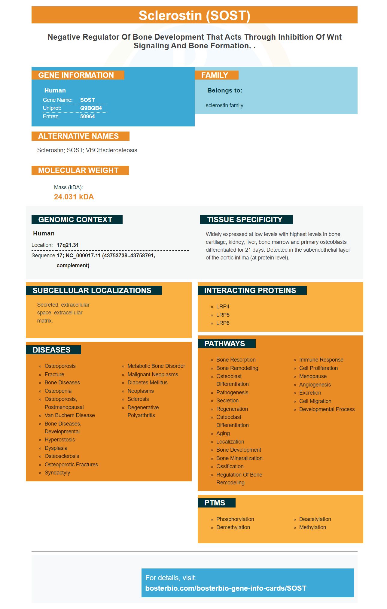
Human | |
---|---|
Gene Name: | SOST |
Uniprot: | Q9BQB4 |
Entrez: | 50964 |
Belongs to: |
---|
sclerostin family |
sclerostin; SOST; VBCHsclerosteosis
Mass (kDA):
24.031 kDA
Human | |
---|---|
Location: | 17q21.31 |
Sequence: | 17; NC_000017.11 (43753738..43758791, complement) |
Widely expressed at low levels with highest levels in bone, cartilage, kidney, liver, bone marrow and primary osteoblasts differentiated for 21 days. Detected in the subendothelial layer of the aortic intima (at protein level).
Secreted, extracellular space, extracellular matrix.
You're not the only one who has ever thought about Sclerostin and its anti-anabolic properties. This article will explore the functions and effects of this biomarker in your studies. This article is an essential resource for biomedical researchers , researchers, and scientists. It will also outline how it can be used to aid you in making better research choices. You can begin using Sclerostin right now.
The skeletal protein Sclerostin is a key component of the bone-vascular axis but its function is not fully understood. However, it appears to play a part in the regulation of fat production and differentiation of Adipocytes. This article provides a summary of the known functions of Sclerostin. These functions can be understood better by examining the molecular mechanisms that Sclerostin utilizes.
Recent research has revealed that sclerostin is a key part in bone homeostasis. The deletion of the Sost gene results in an increase in vertebral BMD in mice. Its ability to neutralize Sost has been proven to be effective in the treatment of osteoporosis. Sclerostin is a key player in osteoporosis, as well as other bone disorders caused by the inability to regulate sclerostin's expression. In human clinical trials, neutralizing antibodies to sclerostin have been used to treat bone disorders.
The mechanism that drives the antagonistic role of Sclerostin in the formation of bone is not fully understood but it is believed to involve the Wnt and BMP signaling pathways. The protein is expressed in osteocytes and a small percentage of chondrocytes. This is believed to reduce the bone-forming ability of osteoblasts. Although the mechanism that causes the antianabolic properties of Sclerostin isn't well understood, it is apparent that sclerostin hinders bone formation by blocking the Wnt signaling pathway.
The SOST gene encodes the protein sclerostin. It was identified through the analysis of genetic linkage in patients with Van Buchem and sclerosteosis. Bone contains many regulatory components that regulate Sclerostin. These regulatory elements tightly control Sost transcription within bone. Therefore, mice lacking LRP5 show an increase in sclerostin.
Although it is unclear what role serum Sclerostin plays however it could be a valuable marker for diagnosing bone disorders and diabetes-related bone disease. More research is needed to determine whether Serum Sclerostin may play a role in other cancers that cause loss of bone. These questions may be answered by the discovery of monoclonal therapeutic antisclerostin antigens. But for now it's an interesting discovery!
A high concentration of esclerostin was associated with an increased risk of cardiovascular events as well as death in patients suffering from CKD. This is because CKD-related cardiovascular disorders are often associated with an altered mineral homeostasis. In addition, the levels of Sclerostin also positively correlate with CKD progression, indicating that it could predict the beginning of cardiovascular events in the CKD population that is not on dialysis.
In this study, we analyzed whether Boster Bio SOST Marker exhibits antianabolic effects in OA Chondrocytes as well as healthy chondrocytes. SOST treatment resulted in an increase in COL2A1 levels and a decrease of b-catenin mRNA level in OA chondrocytes. Furthermore, it inhibited MMP-13, ADAMTS-4 and the expression of p38MAP in healthy chondrocytes. The results were not consistent with the downstream factors.
SOST blocks the Wnt/b-catenin signaling pathway, which has anti-anabolic properties. It decreases the expression of downstream factors, like MMPs and ADAMTSs. Additionally, SOST inhibits IL-1a, an inflammatory cytokine that causes cartilage loss. SOST could delay the progression of OA.
Boster Bio has recently released a new miRNA targeting the SOST marker in Wnt Signalling. The marker, which is part of the ICG-001 protein, inhibits the Wnt signalling pathway, and binds an element-binding protein. In this study we utilized ASCs that were grown in 12-well plate plates, maintained at CO2 and treated with ICG-001 for 2 days.
The SOST marker is utilized in many fields of science that involve manipulation and study of genetic material. Boster Bio's guides provide valuable tips and guidelines to assist you in optimizing your research and get more effective results. Every researcher will encounter issues while completing an experiment. Troubleshooting guides can help you find the root of the problem and increase the value of your data. If you're using Boster Bio as a reference or conducting your own experiments, you'll always be sure to do them under the right conditions.
In a recent study, researchers discovered that miR-218 improves the Wnt pathway through regulation of FGF2 and bIII Tubulin. These results also revealed an interplay between miR-218 and FGF2. The SOST marker pretreatment significantly increases the amount of cells that are double-positive, suggesting that this protein is an essential cofactor in the improvement of Wnt signaling.
This SOST marker is able to monitor the expression of important genes in the Wnt signaling pathway. It controls Wnt3a and the cytoplasmic and nuclear proteins, b–CATENIN and lef1a. The SOST marker also monitors the expression of phosphorylated ZD.
The SOST biomarker kit developed by Boster Bio is a flexible instrument to detect the inflammatory cytokines found in human samples. This kit enhances the sensitivity of ELISA tests up to picogram levels. The immunogen is validated against a panel of 250 human tissues and cell lines. The kit also features high sensitivity and affinity. Technical support is available to answer any questions and help you make the most of your kit.
Sclerostin, a glycoprotein secreted that has a cysteine-like domain. It is part of the DAN family of bone antagonists to morphogenetic proteins. Deficiency of this protein causes an overgrowth of bone, a condition called sclerosteosis. SOST may also be associated with van Buchem disorder that is characterized by a decrease in the expression of the protein.
This ELISA kit analyzes Human SOST levels in cell culture supernatants as well as plasma. The kit comes with 96 well strips. The ELISA kit is able to analyze plasma and serum samples in addition to animal and human serum. The kit is able to detect SOST in human and animal samples. It also contains buffers that contain 0.09 percent sodium azide.
PMID: 11181578 by Balemans W., et al. Increased bone density in sclerosteosis is due to the deficiency of a novel secreted protein (SOST).
PMID: 11179006 by Brunkow M.E., et al. Bone dysplasia sclerosteosis results from loss of the SOST gene product, a novel cystine knot-containing protein.
*More publications can be found for each product on its corresponding product page