This website uses cookies to ensure you get the best experience on our website.
- Table of Contents
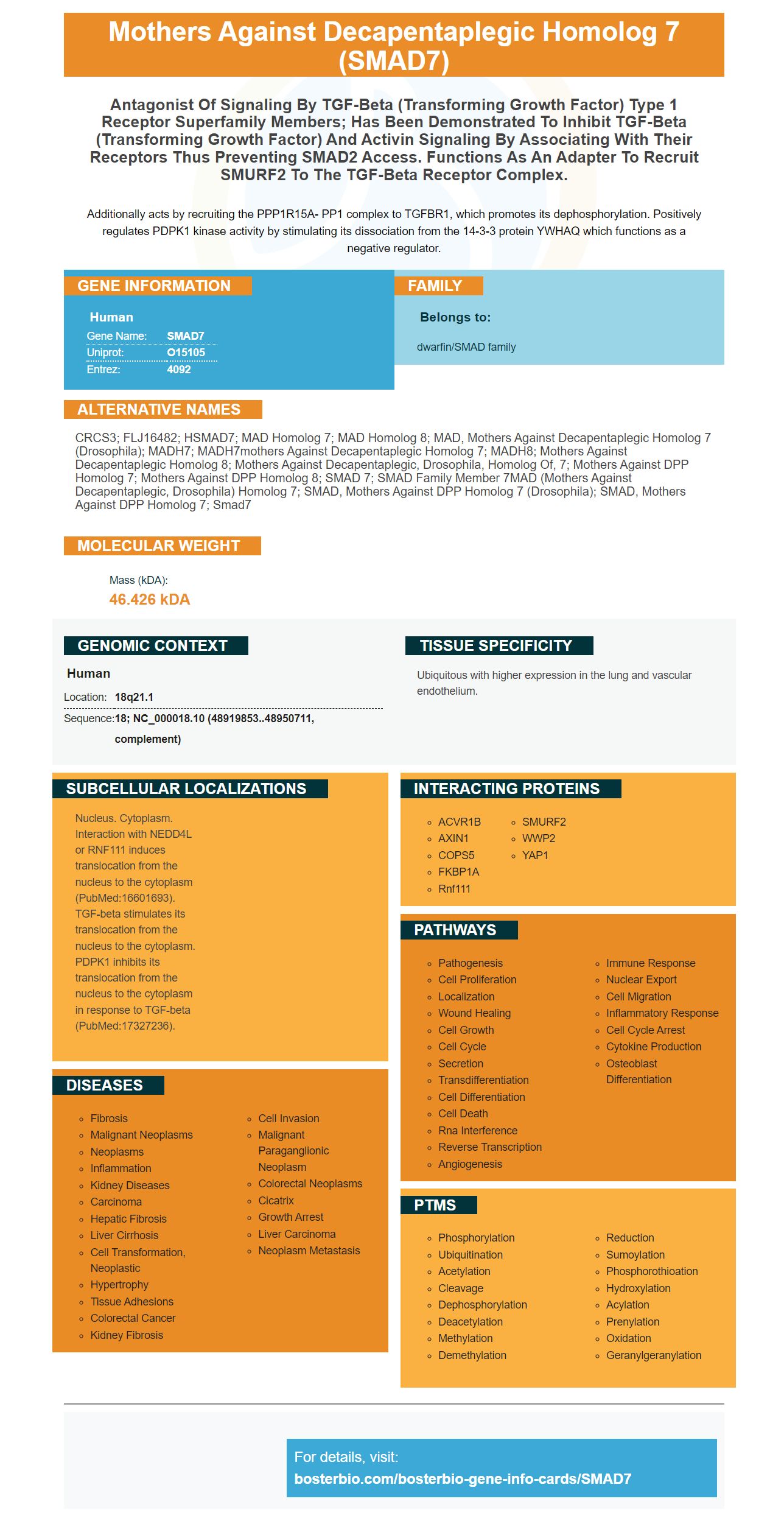
Facts about Mothers against decapentaplegic homolog 7.
Additionally acts by recruiting the PPP1R15A- PP1 complex to TGFBR1, which promotes its dephosphorylation. Positively regulates PDPK1 kinase activity by stimulating its dissociation from the 14-3-3 protein YWHAQ which functions as a negative regulator.
Human | |
---|---|
Gene Name: | SMAD7 |
Uniprot: | O15105 |
Entrez: | 4092 |
Belongs to: |
---|
dwarfin/SMAD family |
CRCS3; FLJ16482; hSMAD7; MAD homolog 7; MAD homolog 8; MAD, mothers against decapentaplegic homolog 7 (Drosophila); MADH7; MADH7mothers against decapentaplegic homolog 7; MADH8; Mothers against decapentaplegic homolog 8; Mothers against decapentaplegic, drosophila, homolog of, 7; Mothers against DPP homolog 7; Mothers against DPP homolog 8; SMAD 7; SMAD family member 7MAD (mothers against decapentaplegic, Drosophila) homolog 7; SMAD, mothers against DPP homolog 7 (Drosophila); SMAD, mothers against DPP homolog 7; Smad7
Mass (kDA):
46.426 kDA
Human | |
---|---|
Location: | 18q21.1 |
Sequence: | 18; NC_000018.10 (48919853..48950711, complement) |
Ubiquitous with higher expression in the lung and vascular endothelium.
Nucleus. Cytoplasm. Interaction with NEDD4L or RNF111 induces translocation from the nucleus to the cytoplasm (PubMed:16601693). TGF-beta stimulates its translocation from the nucleus to the cytoplasm. PDPK1 inhibits its translocation from the nucleus to the cytoplasm in response to TGF-beta (PubMed:17327236).
Biological assays use antibodies to detect the SMAD7 protein. They can be polyclonal or monoclonal. Boster Bio uses mice and rabbits to make their SMAD7 antibodies. The PDPK1 inhibitor blocks SMAD7 translocation and is a potent inhibitor of SMAD7. The antibodies are used for numerous applications in biological assays.
Smad7, an antagonist of TGF-b/BMP signaling is critical for activation STAT3 as well as the downstream pathways of gp130STAT3 and TGFb. It is also implicated in the process of tumorigenesis, and has been found to play a greater role in the bridging of these pathways. Biological assays using anti-Smad7 antibodies suggest its potential use in many research areas.
Many biological assays utilize antibodies against the SMAD7. Monoclonal antibodies to SMAD7 were designed and tested using rabbits and mice as the model species. These antibodies are also available as polyclonal variants. For monoclonal antibodies, Boster Bio uses rabbit and mouse as model organisms for their research.
Smad7 targets PP1c and GADD34 as in addition to TbRI. They also inhibit the function of TGFb by dephosphorylating TbRI and contributing to resistance to UV light. Transfected Mv1Lu cell lines were co-transfected with GADD34-PP1c or 3TP-lux and incubated for 24 hours either without or TGFb-1. The mean + SD of triplicates was used to determine the normalization of Luciferase activity.
Smad7 interacts with TGF-b, which regulates cell potency and functions. It is essential for maintaining pluripotency and self-renewal of mouse embryonic stem cells. Smad7 is responsible for the activation of STAT3 in mouse ESCs. This is a way to promote self-renewal as well as induced pluripotency. Therefore, anti-Smad7 antibody is vital for a variety of biological tests.
Aside from breast cancer, Smad7 has a role in the regulation of TGF-b signaling. Its polyubiquitination is induced through the action of TGF-b and the OTUD1. Its degradation is promoted by RNF12 and ARKADIA as well as ARKADIA. OTUD1 hinders this degrading. Both mutations are linked to decreased survival for patients with breast cancer.
Numerous biochemical tests and immunoassays using Anti-Smad7 antibodies have also proved the effectiveness of paeoniflorin to fight cancer. It stops metastasis-induced by fetal bovine serum as well as increased expression of Smad2. Anti-Smad7 antibodies are used to detect TGF-b1-induced inhibition of Smad7.
These experiments utilized AF6265 as the antibody and Alexa Fluor 488 from Invitrogen. The secondary antibodies were 594 from Invitrogen. In addition to these primary antibodies, we also purchased a number of ELISA kits from Cell Signaling Technology Inc. (Beverly, MA).
SMAD7 is a family of intracellular proteins which are necessary for numerous signaling pathways. There are two kinds that are inhibitory and receptor-regulated. Some have orthologs found in rats and others have canine counterparts. In addition to SMAD7, SMAD6 and SMAD9 have different names in the literature. This boster bio antibody can detect both phospho as mono-Smad3 in an experiment.
Flow Cytometry is a method of analysis that utilizes particles and cells as samples. There are many applications for antibodies that detect SMAD7. These high-affinity primary antibodies from Boster Bio are well-respected and have a track record in the market. The antibodies can be used for a range of purposes and are available in polyclonal as well as monoclonal formats.
HSP72 is a protein that blocks TGFb-induced nuclear translocation and the phosphorylation process of Smad3. It also blocks EMT by preventing Smad3 phosphorylation. HSP72 blocks activation of Smad3 in mice. The knockdown of HSP72 reduced the amount of Smad3 and p-Smad3. This effect was reversed after reintroduction of exogenous HSP72.
Primary antibodies in contrast to secondary antibodies are highly specific and have one target antigen to bind to. They are extremely specific and possess high affinity. They can be used to measure the purity, detect, or purify a specific antigen. They are also available in high-affinity as well as low-affinity versions for immunohistochemistry and ELISA. They are an excellent choice for researchers who need antibodies to study proteins.
A high-specific anti-Smad7 antibodies can target certain targets such as HSP72 and the HSP72P. Both proteins are essential for normal cell function. The antibodies used in immunoprecipitation tests possess excellent affinity for these targets. The antibody can be used in numerous research applications, such as cancer research and drug development. If you want to detect Smad7, you must ensure that your samples contain the protein you're looking for.
HSP72 blocks phosphorylation of Smad3 by securing it in the nucleus. HSP72 inhibits nuclear Smad3 activation when TGF-b1 is exposed to NRK52E cells. It also plays a role in blocking nuclear translocation of p-Smad3. The PBD and NLS interact with each other.
Primer design is a crucial step in determining SMAD7 expression. For the Smad7 gene, the primer sequence comprised of three 'T's was utilized. The primers were designed to work with 307-base-pair (bp) segment. Then, the primers were used in the detection of b-glucuronidase or SMAD7 mRNA.
The SMAD7 proteins also play a role in determining the fate of lymphoid cells. These proteins alter the balance of myeloid cells as well as lymphoid cells. Therefore, studies on humans and other species must concentrate on Smad7 to comprehend the role it plays in lymphoid stem cell development. There are a variety of methods available to measure Smad7 expression in human cells.
Researchers found that mice engineered to express Smad7 had mesenchymal behaviors that are functional. Mice that were engineered to express Ik6 more frequently showed mesenchymal behavior. In this study, mice expressing dominant negative Ikaros 6 (Ik6) were less likely to develop cancer of the colorectal than mice that express Smad7 expression.
Smad7 is a protein that regulates the fate of cancer cells. Studies on molecular biology have revealed that Smad7 is present in cancer cells. The role of Smad7 in cancer cell fate has been previously linked to the fact that it inhibits the EMT process. Therefore, Smad7's overexpression is an underlying mechanism that could regulate the expression of genes in tumors. Further research is required to determine the effects of SMAD7 on cancer cells.
The Smad7 promoter is home to Smad3 and 4 that stimulate the expression of these proteins. The wild type promoter configuration is AP-1's binding site mutated, and Sp1 overexpression is not able to compensate for this. Both DNA binding elements are necessary for transcriptional regulation of Smad7 promoter. If they are affected by mutation they will not be able to compete with the Smad7 mRNA.
The BM of NOD/SCID mice was used to determine human CD34+GFP+ cell lines by using sorting gates. They were then put into a CFU assay using fluorescent microscopy. Retrovirus and fluorescent microscopy were used to determine the SMAD7 CFU–SRCs. Clonogenicity was observed when transduced cells express the promoter Smad7.
PMID: 9215638 by Hayashi H., et al. The MAD-related protein Smad7 associates with the TGFbeta receptor and functions as an antagonist of TGFbeta signaling.
PMID: 9256479 by Topper J.N., et al. Vascular MADs: two novel MAD-related genes selectively inducible by flow in human vascular endothelium.
*More publications can be found for each product on its corresponding product page