This website uses cookies to ensure you get the best experience on our website.
- Table of Contents
2 Citations 7 Q&As
1 Citations
1 Citations
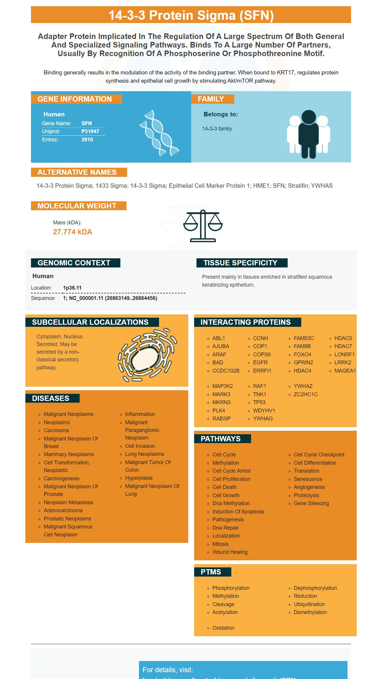
Facts about 14-3-3 protein sigma.
Binding generally results in the modulation of the activity of the binding partner. When bound to KRT17, regulates protein synthesis and epithelial cell growth by stimulating Akt/mTOR pathway.
Human | |
---|---|
Gene Name: | SFN |
Uniprot: | P31947 |
Entrez: | 2810 |
Belongs to: |
---|
14-3-3 family |
14-3-3 protein sigma; 1433 sigma; 14-3-3 sigma; Epithelial cell marker protein 1; HME1; SFN; stratifin; YWHAS
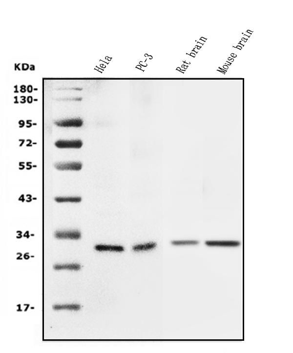
Mass (kDA):
27.774 kDA
Human | |
---|---|
Location: | 1p36.11 |
Sequence: | 1; NC_000001.11 (26863149..26864456) |
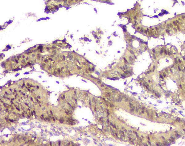
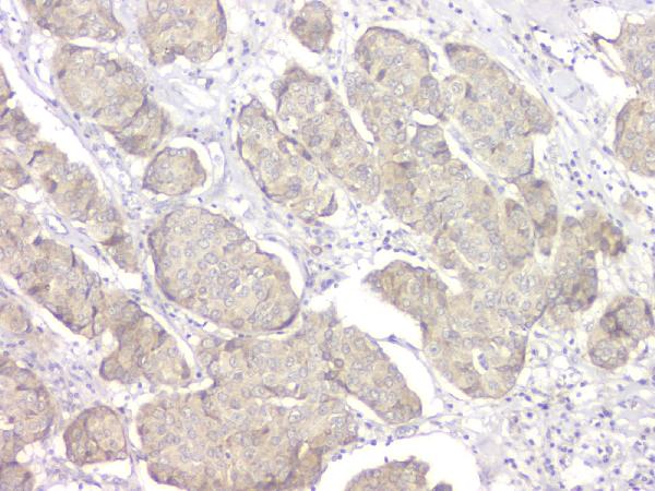
Present mainly in tissues enriched in stratified squamous keratinizing epithelium.
Cytoplasm. Nucleus. Secreted. May be secreted by a non-classical secretory pathway.
This article provides a summary of the benefits Boster Bio's Anti-14-3-3 sigma/SFN Antibody. Continue reading to learn more about how the antibody can be used to verify on WB or IHC and how it can be utilized to treat membranes of the fetus. This article also offers information on how to make the most of Boster's SFN Marker. The ability of this antibody to detect SFN is essential to its effectiveness.
The Boster Bio Anti-14-3-3 sfn antibody recognizes Human, Mouse, and Rat antigens. It has been validated by ICC, IHC, Flow Cytometry and Flow Cytometry. It is available for research, diagnostics and clinical trials. The Antibody can be used for diagnostic and research purposes. It can be stored at -20°C for a period of one year.
The 14-3-3 protein Sigma, also called stratifin, is a commonly-expressed epithelial keratinizing protein that are induced by DNA-damaging agents or radiations such as gamma. It is produced by cells that have been cultured with stratified epithelia. Boster bio Anti-14-3-3 Sigma/SFN Antibody is available in different formats and is cross-validated with confirmed negative and positive samples.
All eukaryotic organisms have the 14-3-3 protein family. Humans have seven isoforms. It is vital for many cells and helps to facilitate interaction between proteins that regulate. The loss of its natural level is associated with various cancers. This is why antibodies against 14-3-3 can be an effective tool in cancer research.
Although these antibodies have been proven to slow the growth of tumors However, further research is needed. Effective cancer gene therapy will require inducible expression of these proteins. These antibodies are very specific to human cancer cells. They have numerous therapeutic applications. They can be used to identify the molecular mechanisms that 14-3-3s interact with p53.
The use of SFN biomarkers could enhance the outcomes of patients with advanced HCC by detecting the disease early and guiding the switch from SFN to alternative therapies. Numerous studies have demonstrated that markers that predict SFN efficacy have been established. However biomarkers are difficult to determine due to the wide range of patients. In this review, we will discuss some of the benefits of Boster's SFN Marker in cancer treatment.
Multiple assays can be utilized to confirm the specificity and authenticity of an antigen. For instance, we could apply Western blot for the first validation step if we know that the antibody recognizes an antigen that is denatured. If the antibody is able to only stain cells that express the target the target, it will be specific. If, however, the antibody exhibits multiple bands at different molecular weights, this could be a sign of various post-translational changes, breakdown products or splice variations. In these instances it is recommended to conduct further tests.
However this WB method has its limitations. For example, if the antibody is unable to recognize the target of a particular receptor, it may not yield the desired results. If the antibody fails to recognize the target, it does not have sufficient specificity to conduct WB tests. In these cases it is recommended to use an animal that has been knocked out. This method, though not perfect, is very useful in proving the specificity of an antibody.
In addition, antibodies may be tested against cell pellets that have been treated with siRNA to inhibit Stathmin expression. This validation can be used to validate antibodies for IHC or QIF. If the antibodies aren't able to show only one band during their validation, you can confirm them using a larger quantity of samples. This method is suggested to detect antibodies that are specific and possess high affinity.
The degree of validation differs from one product to the next. For instance, manufacturers of antibody should be mindful of the reactivity of their antibodies with the cannabinoid CB1 receptor. This warning should be included in the datasheet for any antibody that crosses AMPK B2.
This review aims to explain how to utilize fetal imaging and the most recent methods to detect premature births. We will be discussing the methods used to determine the thickness of the fetal membrane as well as the ability to detect abnormalities. Knowing what is known and what is not will assist us in designing future studies. Clinicians who use CT or ultrasound to assess the thickness of membranes fetal to the fetus have also found this review helpful.
Previous studies have revealed that the membrane of the fetus displayed significant changes in its microstructure during tests of tensile strength in situ. The direction of load causes collagen fibres to reorient in the direction they are supposed to, resulting in a stronger and stiffer AM. These characteristics and the high collagen content have led researchers to believe that AM is a load-bearing element of the fetal membranes. Numerous studies have looked into the strength and deformation of the AM.
The membranes of the fetus begin to deteriorate in utero and continue to age throughout gestation. The premature senescence of membranes in the fetus can cause dysfunction, such as PROM and pTB. In addition, the membranes must be ruptured prior to birth to ensure proper feto-placental delivery. To achieve this, it is important to be aware about the fetal membranes and the factors that could trigger premature senescence.
The mechanical properties of membranes in fetuses can be assessed between ruptured and non-ruptured tissues of the fetus. The AM has the highest capacity to bear loads and is less than the CH. However, ruptured fetal membranes are more likely to be dislocated than those that have not been damaged. This further compromises integrity of fetal membranes. These findings are encouraging and could suggest that fetal membranes may be a treatment option for cardiovascular and neurological diseases.
A novel method of regulating boster cells' PPARg using SFN markers and ligand-independent ChIP is through the use of massive sequencing as well as a ligand independent ChIP assay. This new method will enable researchers to develop more precise maps of DNA-TF interactions. It is vital to identify the binding locations of TFs to identify gene regulatory networks. It is a heterodimer that cooperates with several tissue-selective factors.
The transcription factor PPARg that regulates the expression of many of target genes. The ligands PPARg has include many natural compounds, including eicosapentaenoic acid and linolenic. L-tyrosine-based ligands, nonsteroidal antiinflammatory medications and thiazolidinediones can also be found.
PPARg, a ligand-activated transcript factor that regulates gene expression binding to response elements of promoters of target genes. It performs a variety of functions, including controlling the expression of genes that influence fat metabolism and weight. It is therefore essential to understand the role played by the PPARg in adipocyte development. The role of regulation of PPARg is crucial in fat metabolism as well as the development of obesity.
These regulatory regions are still unexplored , and may be able to trigger points of mutation or major genomic rearrangements. The use of massively parallel sequencing platforms provides the possibility of identifying mutations and regulatory regions that might not have been previously known. These analyses also allow for an exhaustive search for PPARG gene variants that could lead to a causal link to diseases. This study, along with those reported in previous studies, will shed insight into the mechanism of PPARg regulation.
PPARg also plays a major role in macrophages as well as adipocytes. While previous studies focused on its recruitment into cells, recent technological advancements have opened up new layers of complexity. We are now able to analyze the PPARg-mediated regulation across different cell types with innovative methods of sequencing and high-throughput sequencing. This information can be used to discover the mechanisms by which PPARg stimulates specific cellular processes.
PMID: 1390337 by Prasad G.L., et al. Complementary DNA cloning of a novel epithelial cell marker protein, HME1, that may be down-regulated in neoplastic mammary cells.
PMID: 8515476 by Leffers H., et al. Molecular cloning and expression of the transformation sensitive epithelial marker stratifin. A member of a protein family that has been involved in the protein kinase C signalling pathway.
*More publications can be found for each product on its corresponding product page