This website uses cookies to ensure you get the best experience on our website.
- Table of Contents
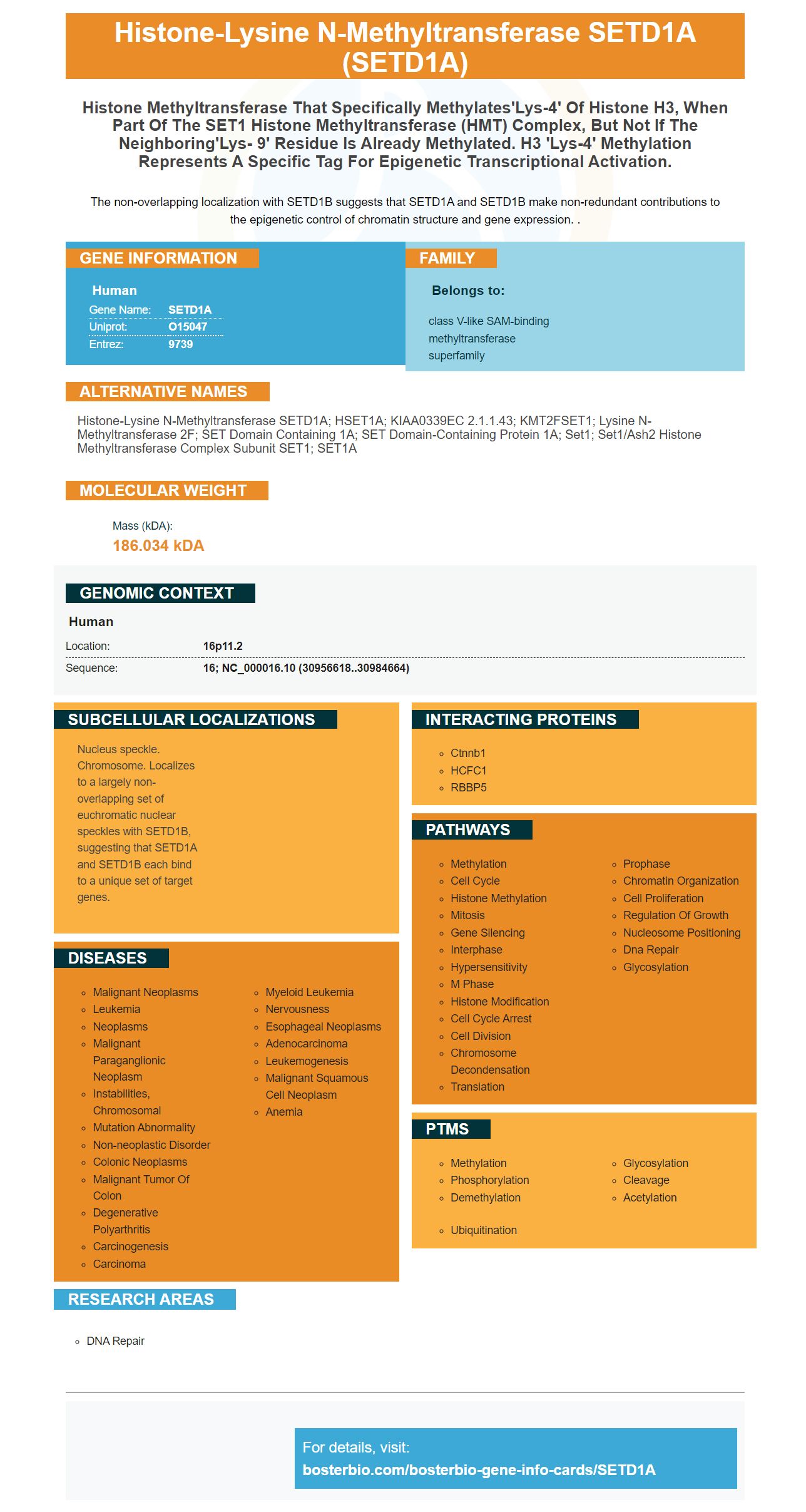
Facts about Histone-lysine N-methyltransferase SETD1A.
The non-overlapping localization with SETD1B suggests that SETD1A and SETD1B make non-redundant contributions to the epigenetic control of chromatin structure and gene expression. .
Human | |
---|---|
Gene Name: | SETD1A |
Uniprot: | O15047 |
Entrez: | 9739 |
Belongs to: |
---|
class V-like SAM-binding methyltransferase superfamily |
histone-lysine N-methyltransferase SETD1A; hSET1A; KIAA0339EC 2.1.1.43; KMT2FSET1; Lysine N-methyltransferase 2F; SET domain containing 1A; SET domain-containing protein 1A; Set1; Set1/Ash2 histone methyltransferase complex subunit SET1; SET1A
Mass (kDA):
186.034 kDA
Human | |
---|---|
Location: | 16p11.2 |
Sequence: | 16; NC_000016.10 (30956618..30984664) |
Nucleus speckle. Chromosome. Localizes to a largely non-overlapping set of euchromatic nuclear speckles with SETD1B, suggesting that SETD1A and SETD1B each bind to a unique set of target genes.
Global histone trimethylation of H3K4 is caused by trimethylation of Histone H3K4 in mammalian cell. This article will describe the primary antibodies as well as their research applications. It is important to know the primary properties of this marker in order to be able to use it. Here are some tips for choosing a good antibody:
Six members of the SETD1A protein group encode proteins involved with histone H3K4 trimethylation. The proteins function in the role of methyltransferases and are involved in many aspects cell physiology as well as development. SETD1A regulates global Histone H3K4 trimethylation.
It is unclear how H3K4me3 markings are targeted at genomes. SETD1A has been implicated as a factor in oogenesis. In fact, previous reports have shown that the redistribution H3K4me3 in GV eggs after Setd1b conditional knoout (cKO), has been demonstrated. Setd1b conditional knockout (cKO) oocytes lose H3K4me3 to gene promoters but gain it at CpG rich regions.
The team of researchers involved in this work included Chen, Patty B. Ding and Shuai, GighA!!, Soulard, ValA (c)rie, and Soulard. The discovery was attributed to the SETD1A protein (a methylation factor that regulates global H3K4 methylation), by the researchers.
SETD1A has been implicated in many types of cancers. It was found that in tumor cells, H3K4me3 levels were decreased compared to the non-cancerous mucosa. This finding has important implications on the study of SETD1A as well as its role in cancer progression.
The SETD1A protein regulates the expression of genes involved in cancer. It has been shown that it is a hallmark active gene-promoters. Furthermore, this protein has been associated with numerous cell activities including cancer cell proliferation and metastasis. These changes can contribute to the development and progression of a disease or predict the likelihood of recurrence.
Genetic research has demonstrated that SETD1A insufficient causes a significant shift in the transcriptomic profile for rodents and humans. SETD1A is enriched for synaptic functions, which causes dysregulated geneexpression. The role of SETD1A and neurodegeneration remains elusive.
The study also found that patients with colorectal disease had higher levels of SETD1A than those with lower expression. Interestingly, this gene is also involved in the expression of H3K4-me3 in colon cancer and colorectal cancer. The SETD1A gene is needed to increase H3K4–me3 levels in colorectal and colon cancer.
The study also revealed that EZH2 expression is independent from its methyltransferase activity. The result demonstrates that EZH2 is essential for neuroblastoma survival and functions in cellular differentiation. It has also been implicated for a variety of tumors including prostate cancer.
The protein was extracted from HeLa S3 cells nuclear extracts by separating the splicing machinery from the unspecific RNP. RNAPII was carried out in the presence ATP as well as a nucleosomal tetramer. RNAPII transcription is dependent upon chromatin or splicing.
The SETD1A protein encodes a protein that belongs to the histone lysine methyltransferase (HLN) complex. This protein produces mono methylated and dimethylated forms histone H3 at Lys4. Trimethylation of histone H3 at Lys4 generally marks transcription start sites. This protein also has an RNA recognition motif. It belongs the class V-like SAM–binding methyltransferase Superfamily. The 16p11.2 address is where the SETD1A Gene is located.
The SETD1A antigen is part of the Boster Bio Picoband(tm) catalogue. It is tested in ELISA, immunohistochemistry, and ELISA. It reacts with Rat, Mouse, and Human. It is well-suited for many diagnostic and research applications. It is available for purchase at tebu.bio. These products can be purchased worldwide.
The retinal development is dependent on the SETD1A protein. It is found throughout the retina. SETD1A is an antibody that recognizes nuclei. This allows the detection and identification of glial cells in your retina. It is expressed in three layers: the inner nuclear layer, neuroblastic layer, and the outer nuclear layer. When SETD1A is analyzed in vitro, it can be detected in all three.
The SETD1A protein is expressed in embryonic retina at high levels but disappears in postnatal stages. Ki67 proliferation antigen shows that SETD1A is expressed in post-mitotic and proliferating cells. It is found in amacrine, Ganglion, and Retinogenic cells, but not bipolar cells. In vitro SETD1A signals overlapped DAPI.
Boster Bio's SETD1A antibody titer for primary antibodies is highly sensitive, specific, and quantitative. Primary antibodies are made from the host's immune systems. The building blocks of these antibodies are called complementarity-determining regions. These topics are complex and are discussed in Wikipedia. Scientists create polyclonal primaries antibodies by immunizing hosts with antigens. These antigens then are extracted from the eggs and sera.
Setd1a, in this case, inhibits the growth or survival of late RPCs. Setd1a regulates expression of two genes: HuC/D (and BRN3B). HuC/D and Setd1a work together to ensure the survival of RPCs. Further research is needed to understand the roles of these genes in CRC. Boster Bio SETD1A, a marker for primary antibody, is an invaluable tool in the field.
The SETD1A marker plays an important role in lung cancer. It regulates the expression of many oncogenes and can provide a novel target for cancer therapy. The gene is found in lung tumors and has a high expression level. Its high levels of expression indicate that it plays an important role in preventing senescence. It could also play an important part in tumor progression.
One study showed that SETD1A inhibited the survival and growth of retinal tissues by increasing the expression levels of the epithelial characteristic gene CDh2. EMT was inhibited in lung cancer patients by SETD1A downregulation. This suggests a possible connection between SETD1A transcription and lung cancer metastasis. Researchers are interested in further studies to determine the best way to use the marker for identifying the source of lung carcinoma.
The SETD1A gene is expressed in both spermatozoa and human embryonic stem cells. Both genes play a critical role in regulating cellular ageing. A depletion in SETD1A results in a decrease of SKP2 expression. This causes a senescent state. SETD1AKD cells show reduced ss-gal staining. SETD1A depleted cells are also known to have extensive mitotic defects and genetic instability.
In addition to the SETD1A gene, Setd1b is required for the survival of neural and epiblast stem cells. The loss of this gene can cause profound learning impairments. SETD1b dependent epigenetic drugs that can restore H3K4me3 peakwidth should be tested. This is important because it is associated with learning behavior and the development of brain tissue. SETD1B-deficient mice might be useful in the development new treatment strategies for cognition.
The SETD1A genes are also involved in mitosis as well as DNA damage repair. The cell cycle competence is linked to SETD1A amplification, which facilitates cell differentiation. For instance, SETD1A knockdown cells exhibit decreased levels of activated b-catenin and the cell cycle protein PCNA. SETD1A knockdown mice had significant reductions in AKT1 levels and phosphorylated STAT3 levels.
The regulation of cell survival and proliferation is also affected by the methylation H3K4 chromatin markers. SETD1A is a significant KMT within the TSS region. Researchers have identified a number of enriched and related GO gene set in SETD1A dependent and SETD1A independent cell lines. This gene will require further research.
This study used a rabbit polyclonal anti-SETD1A antibody that binds directly to the SETD1A protein. The anti-SETD1A antibody used in this study was diluted at a 1:000 dilution to detect Kmt2a/Setd1a. The mRNA extracted from the brains were then resuspended into lysis buffer containing 75mM NaCl 50mM HEPES and 10mM sodium orthovanadate. After being resuspended with lysis buffer, cells could be lysed using a 21 gauge needle. A sample was also taken from the lysates.
PMID: 12670868 by Wysocka J., et al. Human Sin3 deacetylase and trithorax-related Set1/Ash2 histone H3-K4 methyltransferase are tethered together selectively by the cell- proliferation factor HCF-1.
PMID: 16253997 by Lee J.-H., et al. CpG-binding protein (CXXC finger protein 1) is a component of the mammalian Set1 histone H3-Lys4 methyltransferase complex, the analogue of the yeast Set1/COMPASS complex.