This website uses cookies to ensure you get the best experience on our website.
- Table of Contents
1 Citations 1 Q&As
2 Citations 2 Q&As
2 Citations 7 Q&As
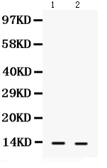
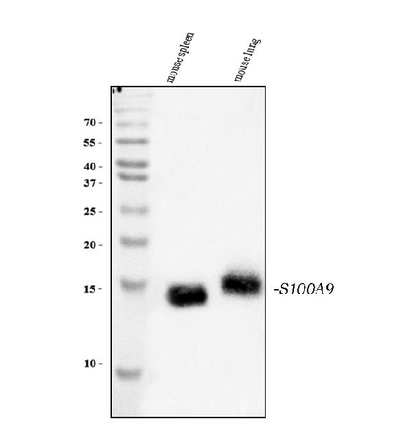
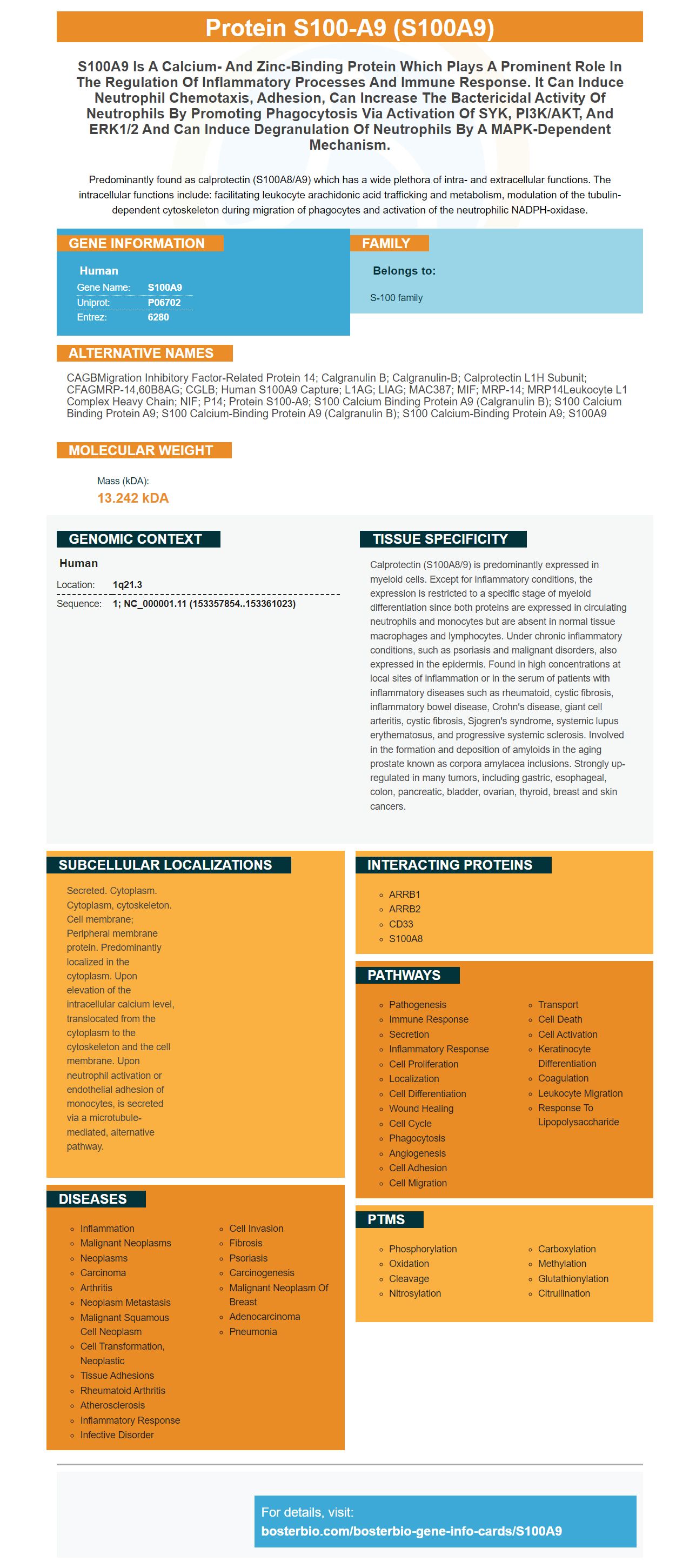
Facts about Protein S100-A9.
Predominantly found as calprotectin (S100A8/A9) which has a wide plethora of intra- and extracellular functions. The intracellular functions include: facilitating leukocyte arachidonic acid trafficking and metabolism, modulation of the tubulin-dependent cytoskeleton during migration of phagocytes and activation of the neutrophilic NADPH-oxidase.
Human | |
---|---|
Gene Name: | S100A9 |
Uniprot: | P06702 |
Entrez: | 6280 |
Belongs to: |
---|
S-100 family |
CAGBMigration inhibitory factor-related protein 14; Calgranulin B; calgranulin-B; Calprotectin L1H subunit; CFAGMRP-14,60B8AG; CGLB; Human S100A9 Capture; L1AG; LIAG; MAC387; MIF; MRP-14; MRP14Leukocyte L1 complex heavy chain; NIF; P14; protein S100-A9; S100 calcium binding protein A9 (calgranulin B); S100 calcium binding protein A9; S100 calcium-binding protein A9 (calgranulin B); S100 calcium-binding protein A9; S100A9
Mass (kDA):
13.242 kDA
Human | |
---|---|
Location: | 1q21.3 |
Sequence: | 1; NC_000001.11 (153357854..153361023) |
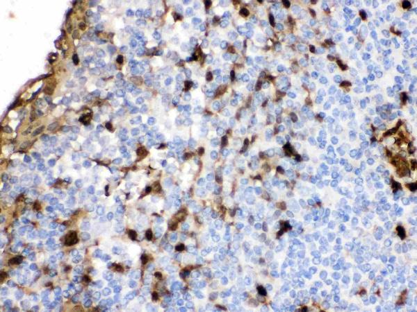
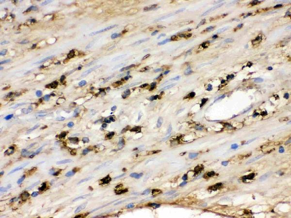
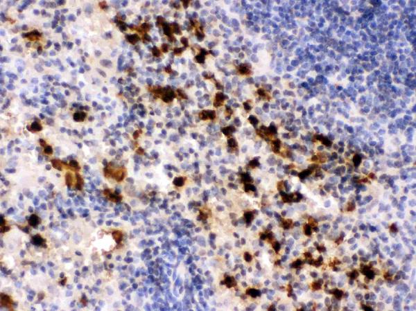
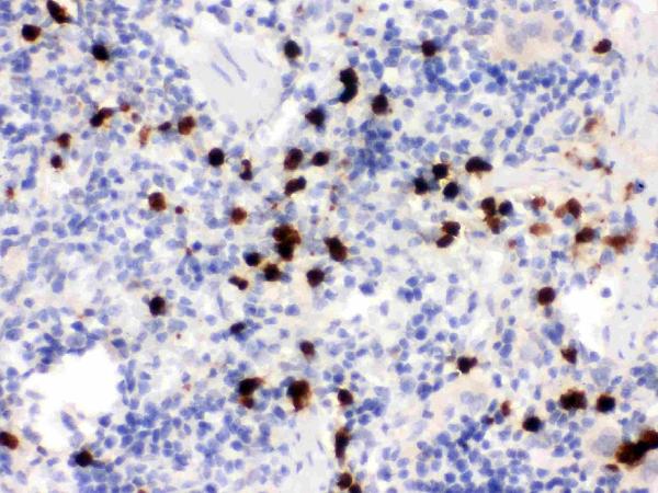
Calprotectin (S100A8/9) is predominantly expressed in myeloid cells. Except for inflammatory conditions, the expression is restricted to a specific stage of myeloid differentiation since both proteins are expressed in circulating neutrophils and monocytes but are absent in normal tissue macrophages and lymphocytes. Under chronic inflammatory conditions, such as psoriasis and malignant disorders, also expressed in the epidermis. Found in high concentrations at local sites of inflammation or in the serum of patients with inflammatory diseases such as rheumatoid, cystic fibrosis, inflammatory bowel disease, Crohn's disease, giant cell arteritis, cystic fibrosis, Sjogren's syndrome, systemic lupus erythematosus, and progressive systemic sclerosis. Involved in the formation and deposition of amyloids in the aging prostate known as corpora amylacea inclusions. Strongly up-regulated in many tumors, including gastric, esophageal, colon, pancreatic, bladder, ovarian, thyroid, breast and skin cancers.
Secreted. Cytoplasm. Cytoplasm, cytoskeleton. Cell membrane; Peripheral membrane protein. Predominantly localized in the cytoplasm. Upon elevation of the intracellular calcium level, translocated from the cytoplasm to the cytoskeleton and the cell membrane. Upon neutrophil activation or endothelial adhesion of monocytes, is secreted via a microtubule-mediated, alternative pathway.
There are many biological assays that utilize antibodies to detect Protein S100A8. These antibodies are either monoclonal or polyclonal and react with Protein S100-A8 across a broad range of animal samples. Boster Bio develops Protein S100A8 antibodies for use in research. Antibodies against S100A8 are beneficial in a variety applications, since it plays a role during inflammation and neutrophil adhesion.
S100A9 is an calcium-bound protein, which bonds to arachidonic acids, components of the cytoskeleton, and the receptor for advanced glycation end products. It is also known to bind to the major fatty acid transporter CD36 and heparin sulfate glycosaminoglycans. Its roles in cell function are not known but include the regulation of microtubule formation and the oxidative potency.
S100A9 is a part of the S100 family calcium-binding proteins. It can be found in a variety of tissues and is expressed by neutrophils, as well as myeloid cells and cancer cells. It is also associated with cancers and is increased in psoriasis. Despite its important roles in cancer cells and inflammation researchers are still trying to discover the precise mechanism of action.
The presence of S100A9 could protect the patient from chronic TB infection. The absence of it can improve Mtb control. In mice that suffer from S100A8/A9 deficiencies the accumulation of neutrophils was reduced. The other myeloid subsets were significantly different from those found in B6 infected mice. In absence of S100A9 neutrophils were found to be more prevalent in the later stages of chronic TB.
Although S100A8/A9 levels in serum were increased during ATB These markers did not rise during COPD or other chronic lung diseases. They could, however, serve as a diagnostic biomarker to detect TB in low-resource environments. This makes them effective in triage. Positive results from biomarkers are able to be followed-up for further TB diagnosis. The levels of S100A8/A9 in the bloodstream are elevated in patients suffering from acute respiratory infections such as influenza. However, further studies are necessary to determine if these levels are elevated in other infections with a bacterial cause.
S100A9, a calcium-bound protein, has binding ligands that are multiple including arachidonic acidsand cytoskeletal elements and the receptor for advanced glycocation end products, Toll - Like Receptor 4 and the major fatty acid carrier CD36. It may block MMP-mediated degradation of the extracellular matrix. It can also block the development of adherent cells in the peritoneum. It also prevents them from phagocytosis. These effects could result from interactions with other extracellular matrix elements and cytoskeletal components, which promote inflammation.
S100A8 and S100A9 are referred to as molecular pattern molecules that cause damage and are involved in the pathogenesis of a variety of diseases. These molecules are produced by neutrophils and have been involved in the development of inflammatory diseases of the central nervous system such as Alzheimer's disease, brain injuries that cause trauma and stroke. It is not clear whether S100A8/A9 plays an important role in the pathogenesis or progression of these illnesses.
S100A8/A9 expression in mice correlates with disease progression. In humans, the protein plays a crucial role in TB immunopathogenesis. In the meantime it has been proven that the combination of S100A8 and S100A9 expression is linked to neutrophil accumulation and improved control of TB. However these findings do not confirm the idea that S100A8/A9 is the sole regulator of the level of inflammation in TB.
Post-translational modification occurs on amino acid side chains, and on protein N and C-termini. They expand the chemical repertoire of 20 amino acids and create an entirely new functional category. Phosphorylation (acylation), lipidation, and phosphorylation are common PTMs. Certain of these modifications are responsible for encouraging protein folding and other functions in the body. Phosphorylation, a redox-active reaction, requires the presence the presence of oxidoreductases (catalytic centre).
The mass-spectrometry-based identification of thousands of PTM sites has allowed scientists to uncover the global cellular significance of several types. Currently, more than 200 types of PTMs are identified. Some modifications are minor chemical changes, like the acetylation process and phosphorylation. Some are more extensive and thorough modifications, such as ubiquitylation. A single modification may be observed on several proteins.
Phosphorylation is a common post-translational modification. It regulates the function and structure of proteins by adding or subtracting amino acid side chains. The most important PTM type, is responsible for regulating protein activity and function. The process of phosphorylation of proteins is reversible. Protein dephosphatases can dephosphorylate proteins. Phosphorylation is vital for cell survival and is a key regulator of many physiological processes.
The S100A9 marker plays a role in cellular apoptosis. It is activated by a variety of factors , including MI, metabolic stress, and reperfusion injury. It regulates cell growth, apoptosis and the process of adipogenesis. Recent research has demonstrated that miR-223 is a regulator of P53/S100A9.
S100A9 plays a part in the development of innate immunity and in inflammatory processes. It is expressed in the kidney's inner core stones, and is not present in the outer matrix. Previous studies have revealed that S100A9 is involved in the nucleation process that initiates crystals. Boonla et al. found that leukocytes that are infiltrated produce an over-expression of S100A8 which is implicated in urolithiasis.
The urinary exosomes of kidney stone sufferers have a higher amount of S100A9 than healthy controls. While the S100 protein was difficult to detect in urine, the expression of S100A8 and S100A9 was elevated in urinary exosomes from kidney stone patients when compared with healthy controls. These findings highlight the importance of S100 proteins in the immune response.
S100A12 is a member of the S100 family and binds with CD36 which is a scavenger class B receptor. The protein directly regulates lipid transport by binding to CD36. A variety of cancer and neurodegenerative ailments have been linked to decreased levels of S100 proteins. Recently, the S100 family has been linked with UCB growth and tumorgenesis. A study found that S100A8 and S100A9 were elevated in UCB patients. Additionally, S100A9 expression was associated with the tumor's grade.
This study demonstrates the ability of an expression signature that is multigene to identify a biomarker that is indicative of UCB. It was created to identify biomarkers in urine and blood samples from UCB patients and healthy groups of control. This multigene signature was found to identify S100A9, S100A12, and p53 in both groups of samples.
The Boster Bio S100A9 anti-S100A9 antibody belongs to the Picoband(tm) catalog. It was tested using ELISA, Flow Cymetry, IF, IHC, and Flow Cymetry. This antibody reacts with both mouse and rat plasma. These results were identical across all species. Sanbio is available to help your lab in analyzing this biomarker. They will be happy to assist you in your research.
This study also revealed that S100A9 is expressed at highest levels in normal bladder tissue and at the lowest levels in tumor tissue. This is in line with previous studies that have identified the gene in squamous cell cancer patients. This may be due to the bladder's immune system being infiltrated by leukocytes. However, further work is required to determine the cell type that is involved in this process.
Boster Bio offers quality reagents and kits for many applications. Boster Bio offers a wide variety of products, including well-known assay kits, buffers, and Lysates. Boster lab produces their antibodies and ELISA kit in-house. They adhere to strict standards to ensure their biological activity. Whether you need an antibody for a basic test or a more complicated study, Boster has the right solution for you.
Established in 1993, Boster Bio specializes in high-specificity and sensitivity antibodies. The Boster lab consistently produces highly tested and validated antibodies and reagents that have been awarded more than 29,000 citations. The Boster lab's antibodies and reagents can be utilized in Flow Cymetry, ELISA and WB. They cover all aspects of neuroscience. Boster Bio's quality assurance ensures that every product performs exactly as it says on the tin.
ELISA kits developed by Boster are extremely sensitive and precise. They can detect a broad array of antigens, proteins and cytokines. With more than 20 years of expertise and the trust of more than 10,000 scientists, Boster offers excellent quality Picokine ELISA kits that speed up the discovery of new scientific knowledge. Picokine ELISA Kits include enough reagents that can be used to conduct up to 96 tests. They can also be tested on various samples.
PMID: 3313057 by Odink K., et al. Two calcium-binding proteins in infiltrate macrophages of rheumatoid arthritis.
PMID: 3405210 by Lagasse E., et al. Cloning and expression of two human genes encoding calcium-binding proteins that are regulated during myeloid differentiation.
*More publications can be found for each product on its corresponding product page