This website uses cookies to ensure you get the best experience on our website.
- Table of Contents
1 Q&As
Facts about 40S ribosomal protein S3.
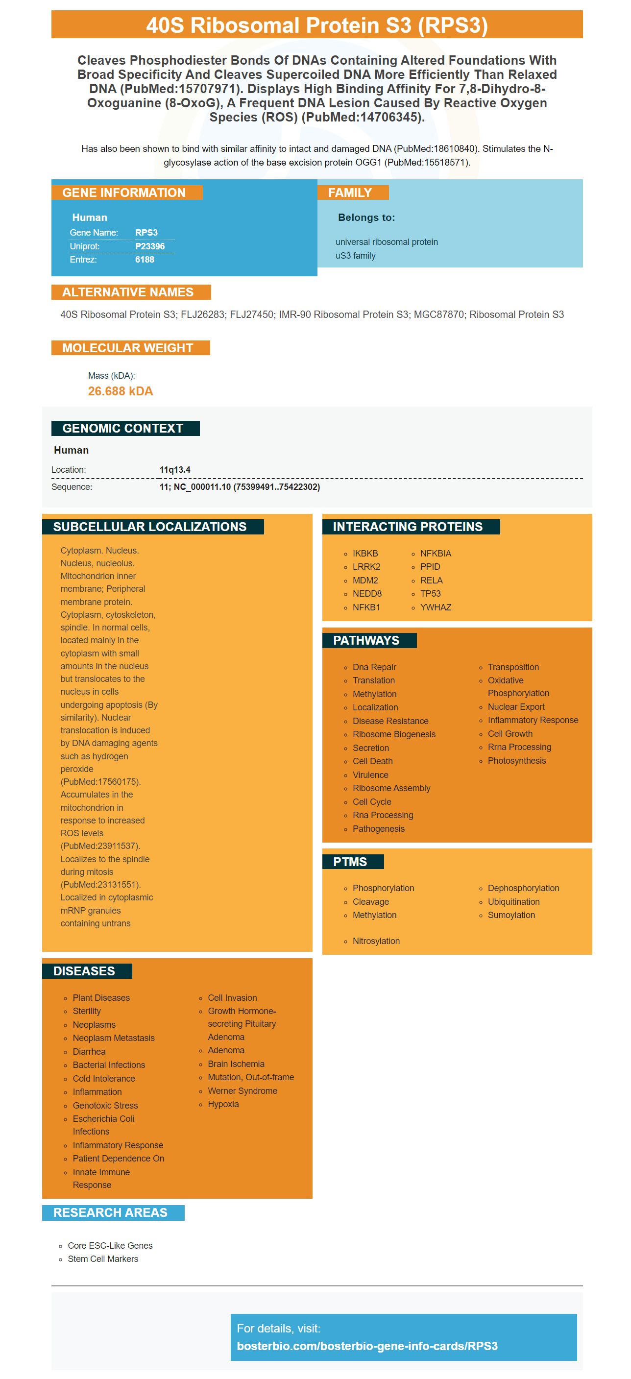
Has also been shown to bind with similar affinity to intact and damaged DNA (PubMed:18610840). Stimulates the N-glycosylase action of the base excision protein OGG1 (PubMed:15518571).
Human | |
---|---|
Gene Name: | RPS3 |
Uniprot: | P23396 |
Entrez: | 6188 |
Belongs to: |
---|
universal ribosomal protein uS3 family |
40S ribosomal protein S3; FLJ26283; FLJ27450; IMR-90 ribosomal protein S3; MGC87870; ribosomal protein S3
Mass (kDA):
26.688 kDA
Human | |
---|---|
Location: | 11q13.4 |
Sequence: | 11; NC_000011.10 (75399491..75422302) |
Cytoplasm. Nucleus. Nucleus, nucleolus. Mitochondrion inner membrane; Peripheral membrane protein. Cytoplasm, cytoskeleton, spindle. In normal cells, located mainly in the cytoplasm with small amounts in the nucleus but translocates to the nucleus in cells undergoing apoptosis (By similarity). Nuclear translocation is induced by DNA damaging agents such as hydrogen peroxide (PubMed:17560175). Accumulates in the mitochondrion in response to increased ROS levels (PubMed:23911537). Localizes to the spindle during mitosis (PubMed:23131551). Localized in cytoplasmic mRNP granules containing untrans
In this article, we'll review Boster Bio's high-affinity primaries antibodies for 40S ribosomal proteins S3 and their diverse applications. Find out more here. Boster Rewards is open to all scientists around the world. We will also pay for the cost of the antibodies and their applications. Let's begin! Find out more here. Learn how Boster Bio products can earn you rewards
Ribosomal protein S3 is one of the subunits of the 40S ribosomal-ribosomal subunit and plays an important role in assessing mitochondrial DNA damage. This protein is a partner with DNA repair proteins, such as uracil-DNA glycosylase in order to increase their activity. It also functions as an endonuclease for repair of DNA damage.
To identify the protein, primary antibodies against the ribosomal subunit can be obtained. Boster Bio has high-affinity primary antibodies that detect rpS3 of diverse species, including mice, humans, and rats. These antibodies are generated by immunizing animals with an peptide synthesized from the lab, followed by purification through peptide affinity chromatography.
Most of the pAbs and mAbs for rpS3 are formulated through conformational mapping. However, a more detailed approach is required to identify the unique amino acid's characteristics. This is crucial since antibodies against rpS3 can be used as biomarkers that aid in the diagnosis and treatment of cancer. If you require a high-affinity antibody against rpS3 in your work, contact Boster Bio today.
A single clone was used to verify the antibody's specificity against RPS3. Western Blotting demonstrated that mAb R2 had higher affinity than the two other antibodies. It recognized the epitope of the rpS3 gene more precisely than mAb R7. However, mAb 8 had lower affinity for rpS3 than mAb 2 suggesting that pAb8 is the best option for your research.
In addition to its flexibility and high affinity affinity, the mAbs play an important role in the management of cancer. Depending on their nature, they can increase the immune response of a patient and act on growth factors for cells. MAbs can be used in conjunction with radioisotopes and toxic substances to enhance the effectiveness of cancer-fighting treatments. Additionally, MAbs can be used in hematopoietic stem cells transplantation.
Primary antibodies are extremely sensitive, high-affinity anti-recombinant proteins and other reagents. For the best results, use an peptide or recombinant proteins diluted to 100 ng/mL in PBST. After the peptide is diluted in PBST and then the primary antibody was added. Then, the membrane was incubated for two hours at 37 degrees Celsius. The primary and secondary antibodies were utilized for immunoassays.
The binding of pAb R2 to rpS3 was determined by competitive ELISA. The binding sites of each Ab were identified based upon their position and their competitive strength with the ligand. A weakly competitive Ab is bound to the antigen target with high affinity, and the pAb R2 binding site was the rpS3 (193-242 aa.
The antibody was created by using HEK293 cells that were differentiated using TLR4-MD2-CD14 ligand. This test demonstrates that RPS3 stimulates DCs in wild-type mouse as well as TLR4 knockout mice. It also was used to create memory T cells, which can be used to stop tumors. The results were quite impressive.
Secretion of the RPS3 marker is a common process in cancer cells and its presence may be a biomarker of malignancy. However, the process of secretion of RPS3 is not well understood. Glycosylation is required for protein secretion, especially Asn165. Additionally, the protein forms a homodimer through interactions between its N-terminal and middle regions. For further study this biomarker is used to identify breast and prostate cancer.
The study revealed three types of markers which could be used for finding the presence of the RPS3 gene in samples. The RPS3 gene is the first marker that was tested. It can be found in all cells. The next step is to determine which of the three types of rps3 markers is the most sensitive and specific. The second type of marker uses RPS3 gene sequences in order to identify cancerous cells. However, its use is still largely unknown.
PMID: 2129557 by Zhang X.T., et al. Isolation of a cDNA encoding human 40S ribosomal protein s3.
PMID: 1712897 by Pogue-Geile K., et al. Ribosomal protein genes are overexpressed in colorectal cancer: isolation of a cDNA clone encoding the human S3 ribosomal protein.