This website uses cookies to ensure you get the best experience on our website.
- Table of Contents
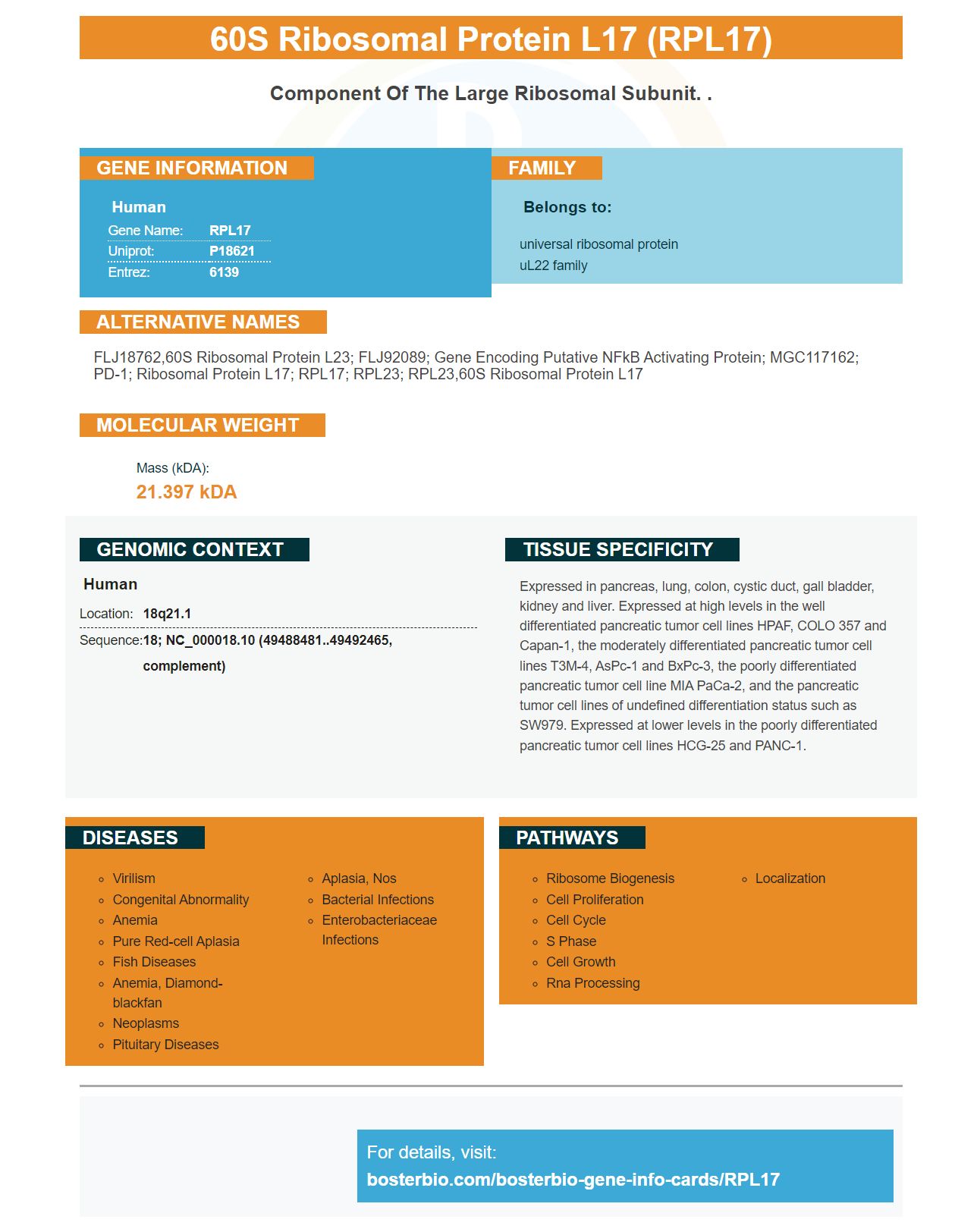
Facts about 60S ribosomal protein L17.
Human | |
---|---|
Gene Name: | RPL17 |
Uniprot: | P18621 |
Entrez: | 6139 |
Belongs to: |
---|
universal ribosomal protein uL22 family |
FLJ18762,60S ribosomal protein L23; FLJ92089; gene encoding putative NFkB activating protein; MGC117162; PD-1; Ribosomal Protein L17; RPL17; RPL23; RPL23,60S ribosomal protein L17
Mass (kDA):
21.397 kDA
Human | |
---|---|
Location: | 18q21.1 |
Sequence: | 18; NC_000018.10 (49488481..49492465, complement) |
Expressed in pancreas, lung, colon, cystic duct, gall bladder, kidney and liver. Expressed at high levels in the well differentiated pancreatic tumor cell lines HPAF, COLO 357 and Capan-1, the moderately differentiated pancreatic tumor cell lines T3M-4, AsPc-1 and BxPc-3, the poorly differentiated pancreatic tumor cell line MIA PaCa-2, and the pancreatic tumor cell lines of undefined differentiation status such as SW979. Expressed at lower levels in the poorly differentiated pancreatic tumor cell lines HCG-25 and PANC-1.
The Boster Bio Anti-60S Ribosomal Protein L17 (RPL17) Antibody has been tested for use in IHC, ICC, and WB. This antibody binds to the human RPL17 gene. It also interacts with the protein that is corresponding to Rat, Mouse, and Human. It is available at a variety of levels and can be used in a variety of applications.
The antisera to Boster's main antibodies and the RP17 marker are extremely effective in the detection of various diseases. The antibodies are tested for reaction with IHC, ICC and WB. Boster's primary antibody and the RPL17 marker are designed to recognize proteins expressed in the cytoplasm of a variety of cells. These antisera also recognize ribosomal protein 3 (a marker for various kinds of cancer). The antisera are highly reactive to different human samples.
To study the expression of a protein blots are typically performed using a monoclonal antibody. Monoclonal antibodies recognize single epitope, while polyclonal primary antibodies are able to recognize multiple epitopes. Western analysis of blots can be done using a variety of imaging systems. The signal intensity of the protein is determined by spectrophotometry.
The concentration of the primary antibody should be diluted as recommended by the manufacturer. The buffer should be diluted to wash the membrane. Do not keep the membrane in the buffer too long , as this can reduce the signal. The membrane is detected using a label antibody, which typically recognizes a specific protein. This enzyme generates signals that match the position of the protein on the membrane. This signal is captured on film and processed in dark rooms.
The proteins of the target are transferred to a hydrophobic membrane, which is then probed using specific antibodies. The proteins of the target are then transferred to the membrane following SDS-PAGE. After the membrane is removed, the primary antibody is reacted with a secondary antibody labeled with an enzyme. The chromogenic and chemiluminescent methods are used to detect the activity of the enzyme bound to it.
The western blot procedure is carried out using a protein extract derived from cells. The extraction of proteins from cell lysates should be done at a lower temperature and protease inhibitors should be employed to prevent denaturing of the proteins. Tissue protein extracts are more complicated and require a mechanical solution. This method also permits greater separation of unfolded as well as not-denatured proteins.
Boster offers high-affinity antibody to the RPL17 marker that can be used in Western Blotting. These antibodies have been mentioned and validated over the last 25 years. They have been tested and proven to be reliable in Western blotting, Immunohistochemistry, and ELISA assays. Researchers utilize boster antibodies for diverse research applications.
MemCode Reversible Protein Stain is a versatile marker that assists in determining the efficiency of transfer and total protein patterns. It is quick and simple to detect. It is also able to be reversed to reduce protein extraction and ensures reproducibility. MemCode staining can be utilized in combination with immunoblotting or other methods like immunoblotting. It can also be utilized in downstream applications.
To determine whether Boster Bio's RPL17 marker improves the transfer of proteins efficiency by membrane staining, first determine the protein concentration in the sample. You can also employ an existing reagent. A regular reagent (200 mM NaOH/20% acetitrile) is enough for this process. Then apply MemCode Reversible stain to nitrocellulose and PVDF membranes. Then, you must wait for at least 20 minutes before washing the membrane. Repeat the staining process on the second or third day.
With this reagent, electroblotted proteins are stained with colloidal gold. The transfected proteins will appear as black lines on grey background. Alkali treatment of the membrane (with 1% KOH and multiple rinses in PBS) improves the sensitivity of the staining reaction. After washing, the membrane is placed in a glass container and the process is repeated three times, lasting 5 min each time.
Boster Bio's RPL17 marker significantly enhances the efficiency of protein transfer using membrane stain. The RPL17 marker is a tool to increase the efficiency of protein transfers in a variety of applications which include electrotransfer. This tool can be used for electrotransfer of 7% SDS-PAGE gels and one that is 10% or 12.5% gel. The RPL17 marker also has the benefit of eliminating trapped proteins from the membrane.
The ECL chemiluminescent detection method is a highly effective tool for biomedical research, and Boster Bio's ECL Chemistry can assist you to carry out this task. This technology has low background, low sensitivity and a long-lasting signal. It is suitable for sensitive biochemical assays and also for measuring tiny amounts of the desired target.
This technology utilizes an emitted chemiluminescent substrate known as HRP. There are many varieties of HRP substrates on the market and each offers different levels of sensitivity. The table below shows the properties of the most popular HRP chemically luminescent substrates. The specificity of each substrate is different depending on the concentration of proteins in the blot. For western blotting, Pierce's ECL substrate is a great choice since it is sensitive enough for detection of proteins that have high levels of phosphorylation.
A blot membrane that contains the protein of interest should be placed in a clear sheet protector of plastic or clear plastic wrap. To eliminate any bubbles or liquid excess, absorbent tissue can be utilized. Place the membrane on the film with Xrays and let it dry for a minute. The initial five - to 30-minute incubation is the time when light emission is at its highest. The process of detection requires an enumeration detector that is chemiluminescent as well as an developer. Finally, fixation is required for a chemiluminescent image.
For a sensitivity-sensitive analysis, Enhanced Chemiluminescence (ECL) is a luminol-based substrate that produces a signal proportional to the concentration of HRP-labeled antibodies. The intensity of the signal is used for measuring the protein concentration. Imaging instruments detect the antigen. The C-DiGit scanner can be used for Western Blots and plate-based assays. However it is also used to study organs that have been excised.
PMID: 2402465 by Mager D.L., et al. A human gene related to the ribosomal protein L23 gene of Halobacterium marismortui.
PMID: 1793733 by Batra S.K., et al. Isolation and characterization of a complementary DNA (PD-1) differentially expressed by human pancreatic ductal cell tumors.