This website uses cookies to ensure you get the best experience on our website.
- Table of Contents
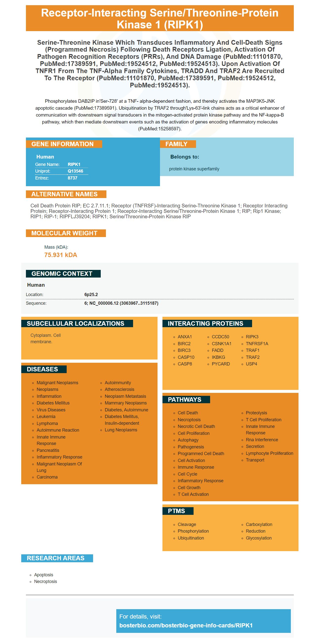
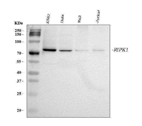
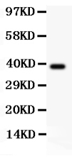
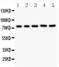
Facts about Receptor-interacting serine/threonine-protein kinase 1.
Phosphorylates DAB2IP in'Ser-728' at a TNF- alpha-dependent fashion, and thereby activates the MAP3K5-JNK apoptotic cascade (PubMed:17389591). Ubiquitination by TRAF2 through'Lys-63'-link chains acts as a critical enhancer of communication with downstream signal transducers in the mitogen-activated protein kinase pathway and the NF-kappa-B pathway, which then mediate downstream events such as the activation of genes encoding inflammatory molecules (PubMed:15258597).
Human | |
---|---|
Gene Name: | RIPK1 |
Uniprot: | Q13546 |
Entrez: | 8737 |
Belongs to: |
---|
protein kinase superfamily |
Cell death protein RIP; EC 2.7.11.1; receptor (TNFRSF)-interacting serine-threonine kinase 1; receptor interacting protein; Receptor-interacting protein 1; receptor-interacting serine/threonine-protein kinase 1; RIP; Rip1 kinase; RIP1; RIP-1; RIPFLJ39204; RIPK1; Serine/threonine-protein kinase RIP
Mass (kDA):
75.931 kDA
Human | |
---|---|
Location: | 6p25.2 |
Sequence: | 6; NC_000006.12 (3063967..3115187) |
Cytoplasm. Cell membrane.
There are countless advantages to using Boster antibodies for your studies. They are validated on multiple platforms and screened with known positive and negative samples, and offer the highest affinity and specificity. Additionally, they reward first reviewers with product credits, and are available for worldwide distribution. Read on to learn about the benefits and features of Boster antibodies. They can help you design your next experiment or research project.
RIPK1 is a kinase that regulates the activity of RIPK3 and thus controls the progression of necroptosis. RIPK1 and RIPK3 are involved in necroptosis signaling and determine the mode of cell death. RIPK1 can be either positively or negatively regulated. It also regulates the activity of caspase-8, a protein that plays a role in necroptosis.
RIPK1 has been shown to suppress autophagy, a process that causes the degradation of proteins in cells. Inflammation is a hallmark of ALS, which affects motor neurons. A recent study by Re et al. demonstrated that RIPK1 inhibits autophagy and apoptosis in a model of ALS. They also discovered that Nec-1 has a protective effect on dopaminergic neurons by suppressing the expression of the autophagy-related protein LC3.
The effects of RIPK1 inhibition on seminiferous tubule regeneration were observed in both wild-type and knockout mice. After three months, 30% of the wild-type seminiferous tubules were empty. The knockout mice, however, were completely unaffected and displayed no loss of cells. Hence, it is likely that RIPK1 inhibits necropsis.
In addition to controlling the autophagy pathway, RIPK1 is also implicated in the development of atherosclerosis. Inflammatory signaling is a major factor in atherosclerotic plaque and is a potential therapeutic target. When activated, RIPK1 inhibits vascular ECs' ability to resist atherosclerosis and develop thrombosis.
In addition to activating NF-kB, RIPK1 kinase inhibits cell death. In addition, RIPK1 kinase is necessary for triggering TNF-dependent ROS in tuberculosis infection. In addition, liver injury has been linked with upregulation of RIPK3 and MLKL. This suggests that necropsis is involved in sterile inflammation.
The kinase RIPK1 controls the process of cell death and inflammation. It inhibits caspase-8-dependent apoptosis and RIPK3/MLKL-dependent necroptosis, but paradoxically induces these modalities. However, the molecular mechanisms underlying the RIPK1 switch between pro-survival and pro-death functions remain poorly understood. Phosphorylation of Ser25 by IKKs inhibits RIPK1 kinase activity and protects cells from the inflammatory effect of TNF.
RIPK1 contains a Glycine-rich loop adjacent to Ser25. This highly conserved sequence motif is believed to help coordinate g-phosphate in ATP during catalysis. Phosphorylation of Ser25 in RIPK1 impairs nucleotide binding, electrostatic repulsion, and structural perturbation of the kinase activation loop. Thus, Ser25 phosphorylation in Boster Bio results in decreased activity of RIPK1 kinase.
In order to identify the effects of phosphatase on Ser25 phosphorylation, cells were transiently transfected with a ribosomal fusion protein. Transfection was performed in petri dishes containing 150 mM NaCl, 10 mM Tris-HCl pH 8, and 1 mM EDTA. Proteins were then isolated by incubation in protein A-Sepharose buffer at 4degC overnight.
In vitro and in vivo, ribosomes containing ROMK1 were labeled with 32Pi. The protein's PKA activity was detected in vivo, but a single mutant did not show a significant reduction in 32Pi incorporation. Moreover, triple mutant ROMK2 showed no PKA-dependent phosphorylation in vitro. Interestingly, phosphorylation of Ser25 did not alter Ba2+-sensitive K+ currents in the cell culture as well.
When ROMK2 is phosphorylated at two sites on the protein, it produces a maximal channel activity. It is important to note that the phosphorylation of all three serine residues is necessary for channel function. The phosphorylation of one of the three serine residues in ROMK2 results in a single site mutant that is 40% lower than the wild-type version.
RIPK1 is a key regulator of RIPK3 oligomerization, a process involved in necroptosis. Its targets are necessary for necroptosis signaling and determine the cell death mode. RIPK3 is also involved in the regulation of FADD-dependent necroptosis. The RIPK1-RIPK3 complex regulates necroptosis, virus-induced inflammation, and programmed necrosis.
RIPK1 is involved in the cell death of many types of cells, including cancer cells. Cell death is also linked to apoptosis. By targeting the RIPK1 marker, researchers hope to overcome some of the therapeutic hurdles in the treatment of inflammatory disease. Here, the authors present the latest findings related to this topic. The authors discuss apoptosis, necroptosis, and caspase inhibition in detail.
The RIPK1 D324A mutation promotes apoptosis and necroptosis, causing embryonic lethality in RIPK1-/ null mice at midgestation. While normal embryonic development is observed in RIPK1-/ null mice, the D324A mutation causes embryonic lethality by E12.5 in RIPK1 null mice. Inhibition of RIPK1 D324A prevents the lethal effects of the mutation, whereas deletion of both alleles prolongs embryonic development.
TNF is another key mediator of cell death. It is also involved in inflammatory processes, as well as in preventing cell death. However, TNF receptors are therapeutic giants. Moreover, Fas/CD95-induced chemokines are important find-me-signaling molecules in apoptotic cells. The RIPK1 marker plays a pivotal role in preventing apoptosis and necroptosis.
Mice with a RIPK1+/D324A founder were used in the study. Mice were used as guinea pigs and RIPK1+/D324A mice were obtained from Dr. Kim Newton at Genentech. All animal procedures were approved by the Institutional Animal Care and Use Committee of Thomas Jefferson University. Once clones of RIPK1+/D324A mice were isolated and genotyped by PCR, and other gene alleles were determined.
The validation of RIPK1 antibodies by Boster is conducted using multiple platforms, including human cells and recombinant proteins. The antibodies are tested for specificity and high affinity against known quantities of recombinant proteins. The antibodies are validated quantitatively using known amounts of recombinant proteins in a panel of 250 tissues. All samples used in the validation process were tested using Boster's proprietary antibody screening methodology.
RIPK1-antibody-derived antisera were validated by staining the mitochondrial membrane protein Tom20. The antiserum reduced expression of downstream proteins such as pNF-KB and pro-IL-1b. In addition, the mouse models expressing the mutant proteins showed decreased Cd-induced AS plaques and increased M2 polarization. The validated antibodies are available for commercial purchase, and data supporting the results of the study can be provided upon reasonable request.
In the validation of RIPK1 antibodies, boster Bio has confirmed their quality. The antibody is part of the Picoband(tm) catalog, and it is suitable for WB and IHC applications. The antibodies are stable in -20°C and 4°C for up to one month. The boster anti-RIPK1 antibody reacts with Human and mouse RIP sequences. To test whether your antibodies are specific, you can purchase blocking peptides.
The TSINGKE laboratory, in Beijing, China, synthesized the PCR primers used in the validation studies. These primers were designed to detect mRNA expression levels of GAPDH, IL-1b, IL-6, TGF-b, HMGB1 and IL-10. Further, TSINGKE also synthesized a RIPK1-antibody against GAPDH, CD86, CD206 (F) and HMGB1.
The RIPK1 gene is involved in several inflammatory and autoimmune diseases. Activation of RIPK1 depends on both the cell type and genetic context of the disease. Certain conditions inhibit RIPK1 activity and increase the likelihood of developing a disease. Inhibition of RIPK1 or other proteins involved in necroptosis is one way to suppress the activation of RIPK1.
While traditional therapies for autoimmune diseases have relied on dampening immune hyperactivity and limiting proliferation of immune cells, RIPK1 mutations may be more effective in determining disease pathogenesis. This may help identify drugs and therapies that target specific mechanisms involved in disease pathology. But there are many challenges in the field of RIPK1 mutations. Here are just some of them. While there are currently no approved treatments, these findings are an important step in the fight against autoimmune diseases.
Some neurodegenerative diseases are associated with dysregulation of RIPK1. Some postmortem human pathological samples reveal a dysregulation of the NF-kB pathway. RIPK1 also has transcriptional targets, such as A20 and cFLIP. Activation of RIPK1 can result in cell death or sustained inflammation. Hence, RIPK1 inhibitors are promising in the fight against MS.
Recent findings suggest that the RIPK1 gene is critical for the progression of sepsis and other inflammatory diseases. Inhibition of RIPK1 inhibits the activity of pro-inflammatory cytokines, preventing the initiation and propagation of uncontrolled cytokine storm. Further, inhibition of the RIPK1 gene inhibits necroptosis, leading to robust protection against sepsis.
PMID: 8612133 by Hsu H., et al. TNF-dependent recruitment of the protein kinase RIP to the TNF receptor-1 signaling complex.
PMID: 7538908 by Stanger B.Z., et al. RIP: a novel protein containing a death domain that interacts with Fas/APO-1 (CD95) in yeast and causes cell death.
*More publications can be found for each product on its corresponding product page