This website uses cookies to ensure you get the best experience on our website.
- Table of Contents
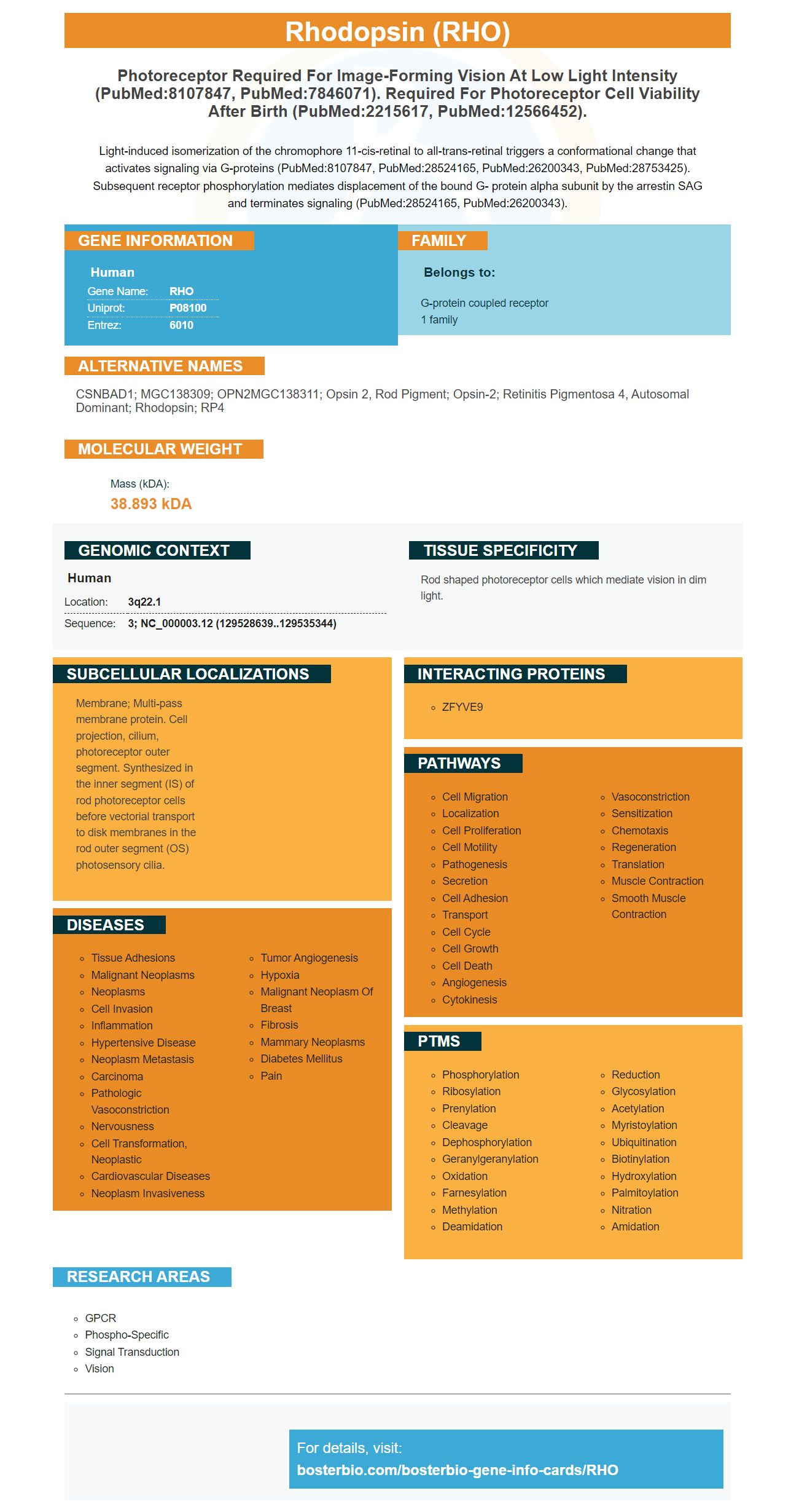
Facts about Rhodopsin.
Light-induced isomerization of the chromophore 11-cis-retinal to all-trans-retinal triggers a conformational change that activates signaling via G-proteins (PubMed:8107847, PubMed:28524165, PubMed:26200343, PubMed:28753425). Subsequent receptor phosphorylation mediates displacement of the bound G- protein alpha subunit by the arrestin SAG and terminates signaling (PubMed:28524165, PubMed:26200343).
Human | |
---|---|
Gene Name: | RHO |
Uniprot: | P08100 |
Entrez: | 6010 |
Belongs to: |
---|
G-protein coupled receptor 1 family |
CSNBAD1; MGC138309; OPN2MGC138311; opsin 2, rod pigment; opsin-2; retinitis pigmentosa 4, autosomal dominant; rhodopsin; RP4
Mass (kDA):
38.893 kDA
Human | |
---|---|
Location: | 3q22.1 |
Sequence: | 3; NC_000003.12 (129528639..129535344) |
Rod shaped photoreceptor cells which mediate vision in dim light.
Membrane; Multi-pass membrane protein. Cell projection, cilium, photoreceptor outer segment. Synthesized in the inner segment (IS) of rod photoreceptor cells before vectorial transport to disk membranes in the rod outer segment (OS) photosensory cilia.
What are the best uses for the RHO marker? This article will cover the clinical applications of antibodies that detect this particular protein in animal samples. This article will focus on the company’s anti-CXCR6 antibody, a G protein–coupled chemokine receiver. Continue reading to learn more. You can also learn about Steven Boster the founder of the company. This company's antibodies for this chemokine receptor are available as boster bio's CXCR4-specific monoclonal antibody.
Many biological assays employ antibodies to detect monoclonal and multiclonal MCM4 proteins. Boster Bio uses mouse and rabbit as models organisms for the development of MCM4 antibody. This antibody binds a peptide that corresponds to the residues around Gly431 in the human MCM4 protein. Boster Bio antibodies that detect MCM4 can be found on the market.
The MCM complex protects and aids in the restarting of the replication process after arrest. It also recruits checkpoint protein after arrest. Its primary function is to maintain chromatin integrity. Antibodies to detect MCM4 could prove to be extremely useful in diagnostic and research. Boster Bio's antibodies to detect MCM4 can be purchased through biotech companies. They are highly specific.
CXCR6 is a G-protein-coupled chemokine receiver that plays divergent roles in homeostasis and inflammation. CXCR6's DRF motif might be an adaptive mechanism. This allows the receptor to maintain its adhesion capability to cells and avoid permanent recruiting of inflammatory. Thus, it may also prevent uncontrolled cancer metastasis.
Cell-specific signaling via CXCR6 is possible. It is well-known that the DRY motif of murine CX3CR1 does little to influence signaling in Jurkat cell cells but increases calcium signaling within THP-1 cells. These differences in signaling could be caused by cell-type-biased signalsing or different G proteins used to signal monocytic cells and lymphocytes.
After just 15 minutes, adhesion to human THP-1 cells was increased by CXCR6 variants DRY and DRF. These results suggest these variants are more efficient than the EV Control in promoting cell attachment. However, these results are not conclusive. These findings are not likely to apply to human cells.
In addition to this, the human class A GPCRs contain two transmembrane chemokines, CXCL3CL1 and CXCR6. CX3CL1 can only be bound by the human CXCR6. The murine CX3CR1Fc protein fusion protein does no interaction with the human CXCR6.
CXCR2 has been shown to have a different affinity than CXCR3. These data indicate that CXCL10/CXCL11 require serine/threonine in their C terminus to be internalized. CXCL11 also depends on transmembrane Helices and requires an additional intracellular loop.
The presence of CXCR6 in the cell culture medium was investigated by transfected BaF3 cells with either mouse or eGFP. The cells were then stained using the rat anti CXCR6 antibodies (catalog # FAB2145C).
CXCR6 shares a similar structure to the class B 7TM receptor. The chemokine receptors, however, are structurally distinct. The chemokine and class A 7TM receptors have similar three-dimensional structures. This makes them a perfect candidate for research and developing new products. Continue reading to learn more about CXCR6.
The RHO marker has a variety of applications. It is a biomarker for diagnosing gastrointestinal bleeding and can be used as a target for therapeutics. However, there are some limitations to the RHO marker. RHO is highly variable in nature, so it is difficult to know what the optimal dose should be. The current status is not clear for RHO-adRP clinical research trials. This review describes the mechanisms of action, current research, development efforts, risks, benefits, and possible outcomes in future trials.
P23H mutation is found in the RHO genes. It is required for normal vision under low-light conditions. There are many types of light receptor cells in the retina, including rods as well as cones. The RHO protein is attached a molecule known as 11-cis retinal. Chemical reactions occur when light hits the molecule. This triggers the formation a signal that is transmitted to brain and interpreted into vision.
The RHO gene has three loops. These loops contain N-glycosylation sites and catalytic site. Additionally, there is a region called lys296. This region attaches directly to the retina. Mutations in the RHO Gene can cause adRP. However only a small percentage of these mutations can cause autosomal-recessive RP. Athanasiou et al. provide an excellent overview of the current research and clinical applications of the RHO marker.
A second candidate gene therapy for RHO-adRP reduces the mRNA of human RHO genes. This shRNA targets the RHO protein in mice and reduces expression of the human RHO gene. The shRNA candidate knocks off the RHO genes in a WT mouse. The injected shRNA knockdown is independent of mutations in the human RHO gene.
You are here because you want to know more about Steven Boster. Steve's public record includes his current and prior addresses, mobile phones numbers, email addresses and known relatives. You can also search for Steve using his age and state. You can also view his criminal history, work history, and if he is divorcing. You can read more about Steve Boster and his family.
Steve Boster was born on November 17, 1944, in Joliet, IL, and passed away in Madison, WI, on June 6, 2022. He was a former retail sales manager and was a member at Concordia Hall in Staunton. He is survived his two daughters, Crystal Boster & Natosha Peck; 6 grandchildren; and 4 brothers, Jack Boster, Sandra Blanton, and Tammy. Many nieces & nephews also survive him.
PMID: 6589631 by Nathans J., et al. Isolation and nucleotide sequence of the gene encoding human rhodopsin.
PMID: 8566799 by Bennett J., et al. Sequence analysis of the 5.34-kb 5' flanking region of the human rhodopsin-encoding gene.