This website uses cookies to ensure you get the best experience on our website.
- Table of Contents
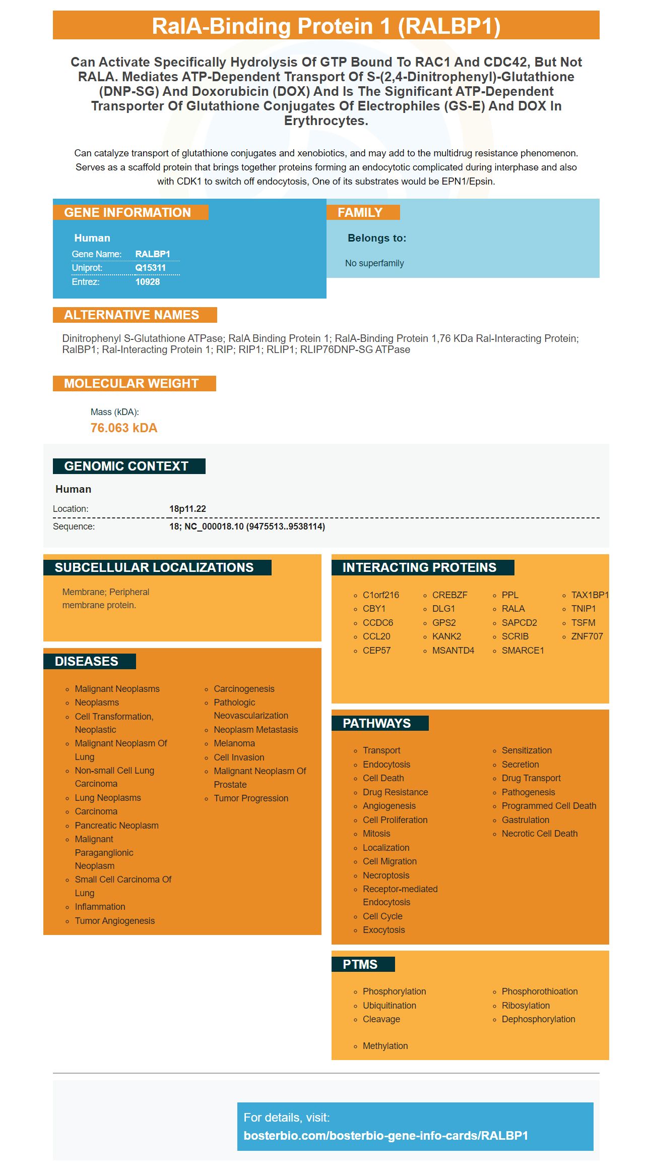
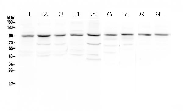
Facts about RalA-binding protein 1.
Can catalyze transport of glutathione conjugates and xenobiotics, and may add to the multidrug resistance phenomenon. Serves as a scaffold protein that brings together proteins forming an endocytotic complicated during interphase and also with CDK1 to switch off endocytosis, One of its substrates would be EPN1/Epsin.
Human | |
---|---|
Gene Name: | RALBP1 |
Uniprot: | Q15311 |
Entrez: | 10928 |
Belongs to: |
---|
No superfamily |
Dinitrophenyl S-glutathione ATPase; ralA binding protein 1; ralA-binding protein 1,76 kDa Ral-interacting protein; RalBP1; Ral-interacting protein 1; RIP; RIP1; RLIP1; RLIP76DNP-SG ATPase
Mass (kDA):
76.063 kDA
Human | |
---|---|
Location: | 18p11.22 |
Sequence: | 18; NC_000018.10 (9475513..9538114) |
Membrane; Peripheral membrane protein.
This article will provide the most crucial details about the RALBP1 marker and its role in mitosis and adipocyte differentiation and many other pertinent topics. We will also explore the most interesting uses of the RALBP1 marker in research. It is important to understand how this marker is produced and used in order to make the most of it. We will go over the various kinds of antibodies and reagents that can be used to create this marker.
The RALBP1 gene encodes a multifunctional GTPase that regulates anchorage-independent membrane raft exocytosis and growth signaling. This gene is a downstream target of the CDC42/RalBP1 signaling pathway and is required for anchorage-independent proliferation in transformed cells. Additionally, it aids in stabilization and the extension of the intracellular bridge during mitosis.
The RALBP1 anti-apoptotic protein is a crucial protein that is found in a variety of cancer cells but it is not required for non-malignant cells. In addition, the loss of RALBP1 causes complete and durable responses in tumor models without leading to harm or weight increase. Ras and Hsf1 also are related to RALBP1.
During mitosis, RalBP1 is located in the cortex of HeLa cells, and to the actin contractile rings during anaphase and the telophase. Western blots were made to determine whether RalBP1 is overexpressed and linked with mitotic spindle and centrosome. The signal in the 95 kDa band was reduced due to the presence of anti-RALBP1 antibody.
The RLIP/RalBP1 enzyme functions as an endocytic partner scaffold as well as a sensitizer to the Epsin phosphorylation reactions. Blocking Ral signaling can have an effect on Epsin phosphorylation. This suggests a function of RLIP in mitosis. During mitosis RLIP76 associates with the active cyclin B1-CDK1 molecule. It also interacts with various endocytotic proteins, such as Numb, AP2, and phosphorylated epsin.
This protein is also necessary for mitochondrial recruitment. RalA, RalBP1, and RalBP1 are phosphorylates on S616 during mitosis. RalBP1 also functions as a mitotic kinase. It also facilitates Drp1's recruitment to mitochondria. These two proteins cooperate to ensure proper segregation of mitochondria. These proteins also work in tandem in order to promote cell viability.
There are many signaling proteins that regulate vesicular traffic and RALBP1 is one of them. This research gives an understanding of the molecular process of RALBP1's effect pleiotropic. Scientists have been able understand the structure of RALBP1 and p19-VHL. In the process of forming the blebbishead for instance p19VHL is involved in the interaction with RalBP1.
RalA is co-reactive with Aurora A, however, the Aurora A phosphorylation has yet to be determined. The study also utilized HeLa cells that are synchronized easily to observe the mitotic mitochondrial dynamics. The study demonstrated that the mutant for RalAS194AA reduced the rate of phosphorylation however not ralA. The knockdown of RalA was reversed by RalBP1-resistant RalAS194 antibodies.
We obtained the GST-RalBP1 expression Virus and DNPSG-affinity reagents to conduct the research. RalBP1 expression plasmids encoding RLIP76 have been previously described (15,29). RNA-seq testing was conducted using 25ng of GST–Cdk1.
Overexpression of RalBP1 in human erythroleukemia K562 cells has resulted in a drug resistant phenotype. The function of this receptor was diminished, which resulted in decreased overall cell drug accumulation and an increase in efflux. The specificity of the resistance was also confirmed by anti-RALBP1 IgG antibodies that bind to epitopes of the cell's surface and block transport activity.
RLIP76 was first identified as the first effector protein of the Ral-GTPase pathway. Ral is a major regulator of cell motility and membrane remodelling. It also regulates endocytosis and actin cytoskeleton remodeling. Furthermore, Ral is a key regulator of metastasis and tumor invasion. It is involved in apoptosis.
RalA and RALBP1 are two of the most prominent proteins that regulate the transport of glucose in the adipocytes. Both proteins are required for glucose transport and are involved in insulin signaling. These proteins in cells could control their functions through coordination. Glut4 and vesicle targets. Molecular models suggest that these proteins could function in a cooperative manner and coordinate the transport of glucose within adipocytes.
Insulin triggers RalA activation in a dose- and time-dependent way. Insulin-stimulated RalA activation is maximum at 5 minutes. A PI 3-kinase inhibitor, wortmannin, blocks insulin-stimulated Glut4 translocation. This suggests that insulin regulates Glut4 localization and trafficking. It is not clear whether RalA regulates the transport of Glut4.
The expression of RALBP1 genes is associated with glucose metabolism and insulin resistance. Researchers have identified 1153 genes that have high and low expression in HPA tissue. The expression pattern of these genes indicates changes in the metabolism of glucose in adipose tissues. The RALBP1 gene is involved with the metabolism of glucose and is a key factor in the regulation of adipocyte differentiation. Furthermore, it is possibly an intervention for obesity.
Numerous studies have proven that LY6CCD9-PDGFRb+ cell lines can serve as precursors for adipocytes. Furthermore, these cells change spontaneously in confluence culture. Friedman and coworkers have found that the LY6C-CD9-PDGFRb+ cells have a higher in FIPs. Further research is needed to determine the precise role this gene plays in the regulation of adipocyte development.
FIPs are a group of transcription factors that are activated by inflammatory signaling. They may act as transcriptional inhibitors against expression of inflammatory chemicals, thus decreasing cell oxidative stress and Apoptosis. Further research is needed to establish whether RALBP1 plays an important role in controlling adipocyte differentiation. There are many uncertainties regarding the role of this gene as a regulator of WAT remodelling in living.
PDGFRb is a marker for cells which has been demonstrated to regulate adipocyte differentiation in mice. PDGFRb+ cells have been proven to differentiate into adipocytes in diet-induced obesity. Additionally, de novo adipogenesis is vital for the growth of WAT. PDGFRb+ cells express both Zfp423 and Pparg.
PMID: 7673236 by Jullien-Flores V., et al. Bridging Ral GTPase to Rho pathways. RLIP76, a Ral effector with CDC42/Rac GTPase-activating protein activity.
PMID: 10924126 by Awasthi S., et al. Novel function of human RLIP76: ATP-dependent transport of glutathione conjugates and doxorubicin.