This website uses cookies to ensure you get the best experience on our website.
- Table of Contents

Facts about Ras-related protein Rab-8A.

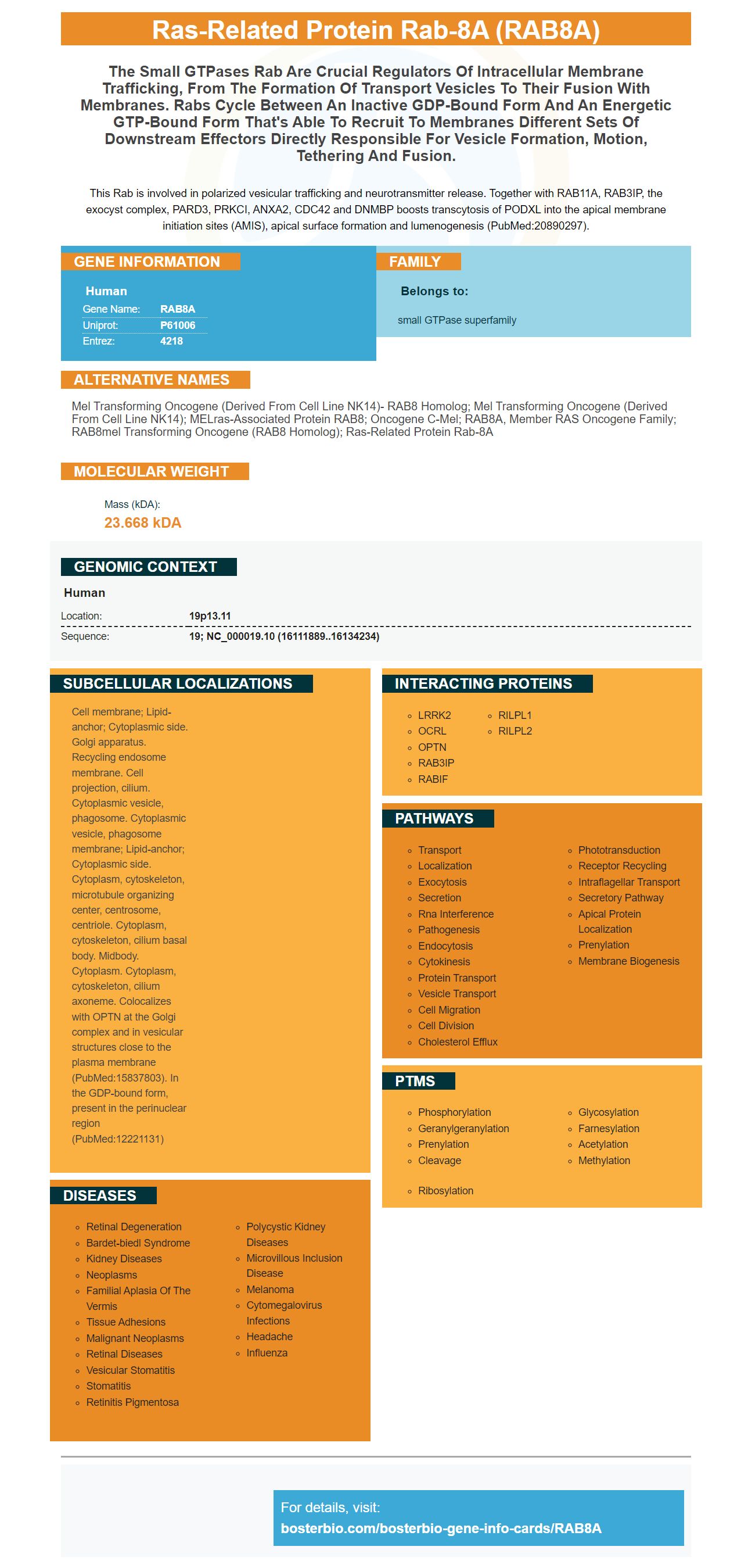
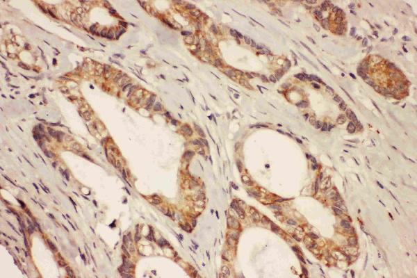
This Rab is involved in polarized vesicular trafficking and neurotransmitter release. Together with RAB11A, RAB3IP, the exocyst complex, PARD3, PRKCI, ANXA2, CDC42 and DNMBP boosts transcytosis of PODXL into the apical membrane initiation sites (AMIS), apical surface formation and lumenogenesis (PubMed:20890297).
| Human | |
|---|---|
| Gene Name: | RAB8A |
| Uniprot: | P61006 |
| Entrez: | 4218 |

| Belongs to: |
|---|
| small GTPase superfamily |

mel transforming oncogene (derived from cell line NK14)- RAB8 homolog; mel transforming oncogene (derived from cell line NK14); MELras-associated protein RAB8; Oncogene c-mel; RAB8A, member RAS oncogene family; RAB8mel transforming oncogene (RAB8 homolog); ras-related protein Rab-8A
Mass (kDA):
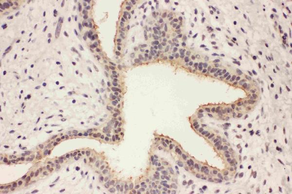
23.668 kDA

| Human | |
|---|---|
| Location: | 19p13.11 |
| Sequence: | 19; NC_000019.10 (16111889..16134234) |
Cell membrane; Lipid-anchor; Cytoplasmic side. Golgi apparatus. Recycling endosome membrane. Cell projection, cilium. Cytoplasmic vesicle, phagosome. Cytoplasmic vesicle, phagosome membrane; Lipid-anchor; Cytoplasmic side. Cytoplasm, cytoskeleton, microtubule organizing center, centrosome, centriole. Cytoplasm, cytoskeleton, cilium basal body. Midbody. Cytoplasm. Cytoplasm, cytoskeleton, cilium axoneme. Colocalizes with OPTN at the Golgi complex and in vesicular structures close to the plasma membrane (PubMed:15837803). In the GDP-bound form, present in the perinuclear region (PubMed:12221131)


PMID: 8294494 by Zahraoui A., et al. A small rab GTPase is distributed in cytoplasmic vesicles in non polarized cells but colocalizes with the tight junction marker ZO-1 in polarized epithelial cells.
PMID: 1886711 by Nimmo E.R., et al. The MEL gene: a new member of the RAB/YPT class of RAS-related genes.
