This website uses cookies to ensure you get the best experience on our website.
- Table of Contents
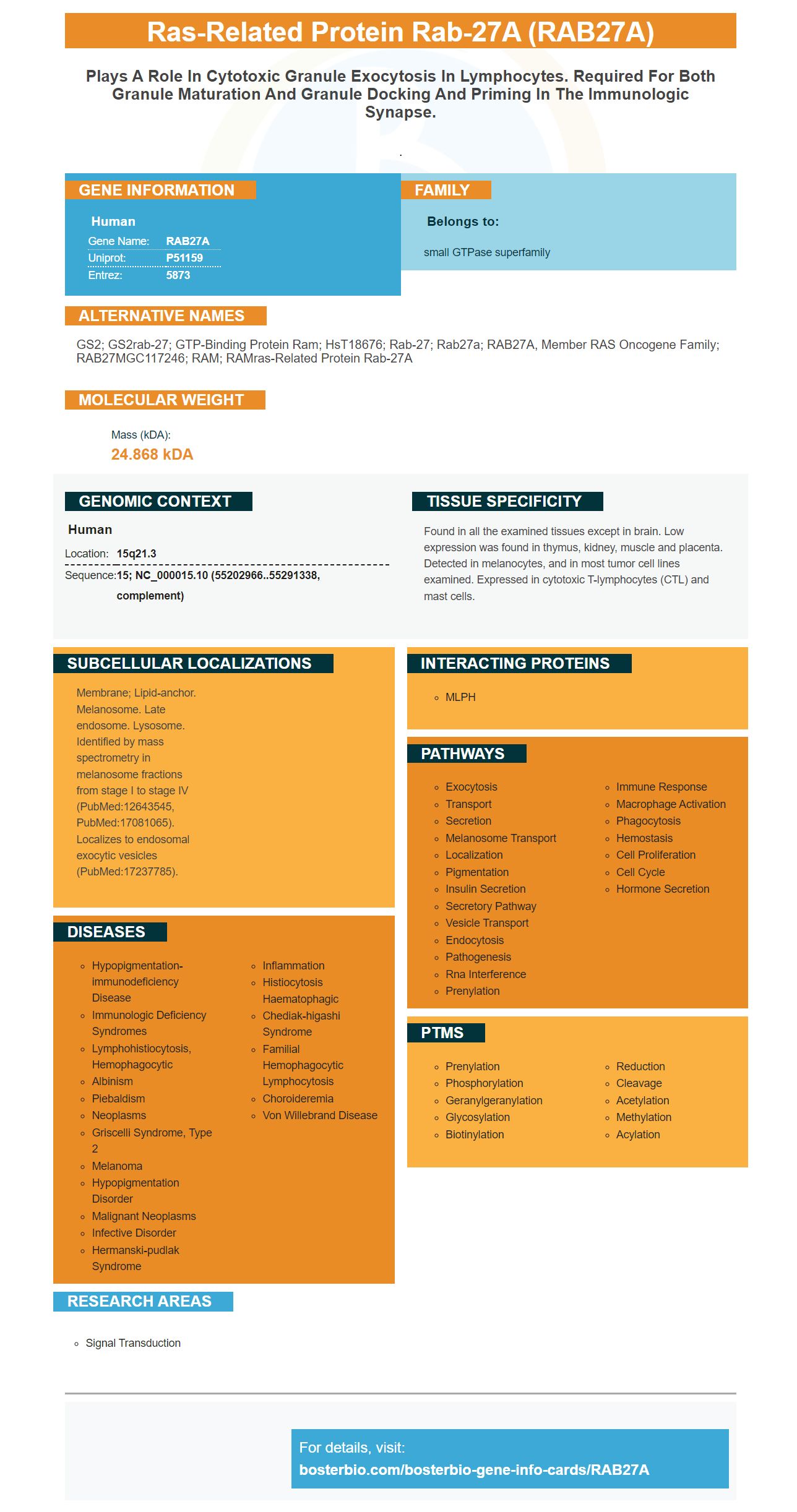
Facts about Ras-related protein Rab-27A.
.
Human | |
---|---|
Gene Name: | RAB27A |
Uniprot: | P51159 |
Entrez: | 5873 |
Belongs to: |
---|
small GTPase superfamily |
GS2; GS2rab-27; GTP-binding protein Ram; HsT18676; Rab-27; Rab27a; RAB27A, member RAS oncogene family; RAB27MGC117246; RAM; RAMras-related protein Rab-27A
Mass (kDA):
24.868 kDA
Human | |
---|---|
Location: | 15q21.3 |
Sequence: | 15; NC_000015.10 (55202966..55291338, complement) |
Found in all the examined tissues except in brain. Low expression was found in thymus, kidney, muscle and placenta. Detected in melanocytes, and in most tumor cell lines examined. Expressed in cytotoxic T-lymphocytes (CTL) and mast cells.
Membrane; Lipid-anchor. Melanosome. Late endosome. Lysosome. Identified by mass spectrometry in melanosome fractions from stage I to stage IV (PubMed:12643545, PubMed:17081065). Localizes to endosomal exocytic vesicles (PubMed:17237785).
In this article, we'll speak about the advantages of RAB27A for Protein Transfer from autoradiography films and membrane staining. We'll also explore how this marker could benefit researchers who need to study specific species or applications. This article is applicable to all scientists worldwide regardless of their the field they are in. Here are some of the best uses for the RAB27A Marker. Read on to learn more!
A membrane stain is required to verify the protein transfer efficiency. This will ensure reliable and high-quality results. The Ponceau S staining solution (0.2% with Ponceau S in 5.5% glacial Acetic acid) proves the efficiency of the transfer of proteins. However, it's not enough to stain the membrane with Ponceau S, as the dyes can interfere with the binding of antibodies to proteins. It is important to select a stain that is easy to remove. Ponceau S is the most frequent stain. It has a limited range of photography and sensitive. In addition, it fades quickly, making documentation difficult.
Picokine(tm), a platform from Boster Corporation, allows for high-sensitivity ELISAs ranging from sensitivity to the picogram level. These kits can be adapted to specific samples and applications. Alternatively, Boster's immunostaining services can help you identify the specific protein you'd like to measure. Boster Bio provides high-quality digital images to help you analyze.
This method requires the preparation of a gel comprising 25 mM Trisbase, 190 mg of glycine and 20% Methanol. Based on the size of protein, thicker gels may cause a lack of transfer since the protein has to travel further to reach the membrane. However, this approach provides another benefit: it can increase the capacity of the gel, which means it will be able to store more proteins in the electroblotted membrane.
Immunoprecipitation with the same antibodies on different membranes can significantly reduce the amount of time needed to carry out the test. Re-probes of proteins can make the method more precise. This technique can cut down on the cost and time required to conduct a protein transfer experiment. The process can be repeated as frequently as necessary. It is a time-saver to have a high-quality antibody that has been tested on multiple targets.
Improved electroblotted membranes can stop proteins from splitting during subsequent incubations. The sensibility of western-blotting also has been improved using 0.5 percent glutaraldehyde for electrophoresis. The supported molecular-matrix electrophoresis however, doesn't require transfer. This method uses the fixation process that is similar to the procedure used for immunoblotting. Fixation is achieved through heating or immersion of the PVDF membrane.
This method is most common for protein transfer it employs both wet and dry methods. A sandwich constructed from an emulsion filter sandwiched between two buffers for transfer is a wet transfer sandwich. The sandwich is then suspended vertically in a transfer tank. This is a fantastic method of transferring protein. It is contingent on the degree to which gel binds to the membrane. Moreover, the sandwich is slower than the other two methods, and it is more susceptible to bubbles between the layers of the sandwich.
Another method to determine the efficiency of transfer of proteins is to determine the molecular mass of the proteins being transferred. Proteins with a lower molecular weight can be detected using a lower-pore membrane, whereas high-molecular-weight proteins like CerP can be labelled with high-pored membranes. It is therefore crucial to choose the proper membrane for your protein.
In a recent study Boster Bio discovered that their RAB27A efficiency of transfer of proteins by autoradography film was about 10 times greater than the original reagent. Rab27a is a membrane-bound, secreted vesicle with similarities in structure to multivesicular exosomes. Although exosome biogenesis remains unanswered, it could play a crucial role in intercellular communication and the development and progression of degenerative and infectious diseases. Rab27a and Rab27b are both involved in docking at the plasma membrane as well as redistribution toward the perinuclear zone and the perinuclear region, while Rab27b has a distinct role in the exosomal pathway.
The Rab27A marker has been developed to aid researchers in targeting specific proteins to prevent cancer metastasis. Its role is to mediate docking and the transport of secretory vesicles to the plasma membrane. Rab27A is essential in the production and transport of chemokines and metalloproteases. These features make Rab27A a great target to disrupt cancer metastasis.
The overexpression of Rab27a has been linked with a poor prognosis in breast cancer. It is believed to be responsible for extensive remodelling through systemic EV release. However, the exact function of Rab27a in pancreatic cancer is not clear. The goal of the present study was to determine if Rab27a influences metastatic properties of pancreatic cancer in the vivo. It is crucial to recognize that Rab27a expression is increased in ER-positive breast cancer, which is more aggressive.
The RAB27A Marker has many potential benefits for the development of drugs. It can aid researchers in identifying novel compounds and confirm targets. Competitive inhibitors have been directed towards the SF4 pocket which is a particular challenge for small molecules. A covalent library could be used to identify Rab27A-targeted small molecule. Researchers now have a instrument to design new drugs that target the Rab27A molecule, which is a RAB27A marker.
PMID: 7592656 by Seabra M.C., et al. Deficient geranylgeranylation of Ram/Rab27 in choroideremia.
PMID: 9066979 by Chen D., et al. Molecular cloning and characterization of rab27a and rab27b, novel human rab proteins shared by melanocytes and platelets.