This website uses cookies to ensure you get the best experience on our website.
- Table of Contents
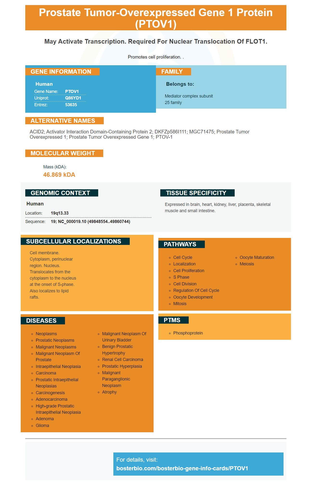
Facts about Prostate tumor-overexpressed gene 1 protein.
Promotes cell proliferation. .
Human | |
---|---|
Gene Name: | PTOV1 |
Uniprot: | Q86YD1 |
Entrez: | 53635 |
Belongs to: |
---|
Mediator complex subunit 25 family |
ACID2; Activator interaction domain-containing protein 2; DKFZp586I111; MGC71475; prostate tumor overexpressed 1; prostate tumor overexpressed gene 1; PTOV-1
Mass (kDA):
46.869 kDA
Human | |
---|---|
Location: | 19q13.33 |
Sequence: | 19; NC_000019.10 (49848554..49860744) |
Expressed in brain, heart, kidney, liver, placenta, skeletal muscle and small intestine.
Cell membrane. Cytoplasm, perinuclear region. Nucleus. Translocates from the cytoplasm to the nucleus at the onset of S-phase. Also localizes to lipid rafts.
If you're looking to find a novel marker for your research, then look no further. Boster Bio offers scientists around the world the chance to submit their results using PTOV1. The PTOV1 proteins is found in cells from the beginning of mitosis through the end. It induces progression throughout the cell cycle. The results you submit may be used for a wide variety of applications.
Recent studies have shown that prostate cancer patients with high levels of PTOV1 expression are more likely to be active proliferators. PTOV1's overexpression correlates to higher Ki67indices, which are indicators of cell proliferation. Moreover, PTOV1 overexpression is associated with higher serum PSA levels. However, more research is needed on its role in prostate tumors.
We used immunostaining as a method to determine if PTOV1 expression is associated with proliferative activitiy. We performed immunostaining by measuring the expression of PTOV1 and Ki67 in tumor samples. The resulting staining Index was a function if the PTOV1 antibody was reacted in the cells. Normal prostate tissue was not stained with PTOV1 (a negative controls), but there was strong staining. However, cytoplasmic expression of PTOV1 occurred in high-grade patients. The tumor sample had low and moderate levels of PTOV1, while low-expression was characterized as a staining score of less that four.
PTOV1 expression in HG pin lesions was also tested. Positive PTOV1 expression was related to the presence of cancer, a study showed. This result was similar when comparing prostate biopsy samples with those who had not been diagnosed with prostate cancer. Patients with high PTOV1 expression are not at greater risk for developing prostate cancer. However, it is best to detect it during a follow up examination.
During the growth phase, PTOV1 is mostly localized to the cytoplasm and partially translocated to the nucleus. These cells were forced into S phase by transfection with chimeric green fluorescent proteins-PTOV1. This led to an increase in the level of cyclinD1 protein and forced them into S phase. The proliferative status of prostate cancer cells is correlated with their biological behavior.
An increase in cell proliferation is associated with PBOV1 expression in prostate cancer. It regulates several key proteins that are involved in the G1/S switch. Moreover, prostate cancer cells that express PBOV1 are more likely not to require anchorage. Hence, prostate cancer cells overexpressed PBOV1 are associated with higher proliferative rates and larger colony sizes.
Transfection of chimeric GFP-PTOV1 with human prostate cancer cell lines was performed using Lipofectamine. After transfection, cells received were fixed in 4% paraformaldehyde. They were then treated using anti-PTOV1 antibody. Confocal microscopy then was used to examine GFP-positive prostate cancer cells. Interestingly, chimeric green fluorescent protein-PTOV1 was transiently transfected with a GFP expression construct containing the nonchimeric GFP.
For each of the five tumor subgroups, a positive p value of PTOV1 was calculated and compared with the serum AFP202 level. Patients with high PTOV1 expression had a lower chance of their tumors recurring. These findings highlight how important it is to monitor prostate cancer using PTOV1.
The nuclear protein PTOV1 regulates many cell processes, including growth, differentiation, proliferation, etc. Its receptor induces entry into the nucleus, where it acts as a transcription factor for a diverse set of target genes. These include the HES1 and HEY1 genes. Interestingly, this protein also acts as a transcriptional repressor on a subset of these targets.
Various diseases are linked with the expression of PTOV1 (Nucleus) including leukemia, breast, and ovarian. Its proliferative and tumourigenic abilities are also linked to its existence in the nucleus. Although it is not yet known what PTOV1 does, it may play a key role in the progression of metastases and tumor growth.
PTOV1 was overexpressed in prostate cancer cells and in the prostate-derived enometriosis cell line (IEN). PTOV1 expression was associated with both high proliferation, and nuclear localization. PTOV1 was also overexpressed in PC cells, which led to an increase in the expression of cyclin D1 cells. It may be useful in the early detection of prostate carcinoma.
A recent study revealed that PTOV1 was found in high numbers in atypical adenomatous Hyperplasia (AAH), a benign proliferative lesion that resembles a low-grade carcinoma. However, PTOV1's localization in the nucleus and its effect on mitosis in PC cells remains elusive.
PTOV1 has also been shown to negatively impact chemotherapy for NSCLC. Depletion of PTOV1 decreases cell survival and enhances the sensitivity of cancer cells to chemotherapy drugs. It also decreases the levels of nuclear factor kappa B. As a result, PTOV1 depletion improves the effectiveness of chemotherapy for NSCLC.
PTOV1 regulates the DNA damage response. It also promotes growth and survival. ATP and dATP binding strongly regulate the protein's expression, activity, and function. However, its phosphorylation by the Cdk1/2 enzymes CREB2 and RRM2 during cell-cycle progression is not reliant on ATP.
During the S-phase of the cell cycle PTOV1 induces the nuclear localization and development of flotillin-1. The HA tags were placed at the amino and carboxy termini for flotillin-1. The HA tag was placed on the amino-carboxy terminus of a truncated version of flotillin-1, which lacks the first 23 amino acids. The protein was detected inside the nucleus cells of synchronized PC-3 cells. It was a strong inducer in the S phase.
In several subgroups LUAD and LUSC, the expression of PTOV1 was statistically significant. In addition, poor overall survival was significantly correlated by PTOV1 expression. Kaplan Meier plotter database also showed that PTOV1 was associated with poor progression survival in LUAD, LUSC and LUSC patients. High levels PTOV1 expression in NSCLC is a poor prognosis indicator.
Moreover, chemotherapy for NSCLC may be affected by the depletion PTOV1. Compared to cells that were not treated, PTOV1-depleted cell lines were more responsive to chemotherapy drugs than untreated ones. They survived better when docetaxel and cisplatin were used. As a result, inhibiting PTOV1 expression enhances the chemosensitivities of NSCLC cells.
PTOV1 is a mitogenic proteins that has been previously shown to be active in prostate cancer cells. Various combinations PTOV1 has been shown to be mitogenic, including those tagged with flotillin-1 (or GFP) proteins. Highly mitogenic is also FLAG-PTOV1. The protein has been shown to enter the nucleus. Transfection of individual PTOV domains reduced their mitogenic capacity, but transfection of full PTOV1 replicates the full effect.
Notch signaling (NRP–Jk) and the Notch complex (notch repressor compound) play a central role when regulating the activity PTOV1. Notch signaling is inhibited by Notch. This is done by increasing PTOV1 nuclear localization. Notch activation results in the disengagement of PTOV1 from HEY1/HES1 promoters. This suggests that PTOV1 may block Notch signaling.
HA-PTOV1 overexpression promotes anchorage-independent growth and invasion in PC-3 cells. In vitro PTOV1 inhibits Notch2 induced invasion. The transfection of PC-3 cells with GST-PTOV1 inhibits Notch2-induced invasion in a cell culture dish. Further studies are needed to identify the exact mechanisms that PTOV1-mediated invasion of cells.
20 primary tumors were found to have HES1 or PTOV1 while 16 lymph node metastases had PTOV1 and BPZ. In BPZ and HGPIN, PTOV1 expression was low, while HEY1 and HES1 showed strong staining. Cancerous cells had a higher cytoplasmic concentration of PTOV1.
PMID: 11313889 by Benedit P., et al. PTOV1, a novel protein overexpressed in prostate cancer containing a new class of protein homology blocks.
PMID: 14657022 by Mittler G., et al. A novel docking site on Mediator is critical for activation by VP16 in mammalian cells.