This website uses cookies to ensure you get the best experience on our website.
- Table of Contents
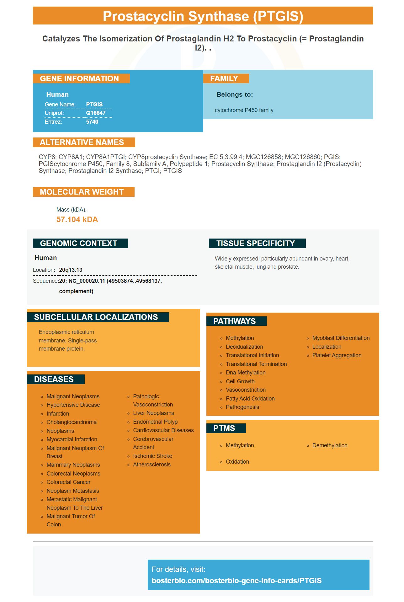
Facts about Prostacyclin synthase.
Human | |
---|---|
Gene Name: | PTGIS |
Uniprot: | Q16647 |
Entrez: | 5740 |
Belongs to: |
---|
cytochrome P450 family |
CYP8; CYP8A1; CYP8A1PTGI; CYP8prostacyclin synthase; EC 5.3.99.4; MGC126858; MGC126860; PGIS; PGIScytochrome P450, family 8, subfamily A, polypeptide 1; Prostacyclin Synthase; prostaglandin I2 (prostacyclin) synthase; Prostaglandin I2 synthase; PTGI; PTGIS
Mass (kDA):
57.104 kDA
Human | |
---|---|
Location: | 20q13.13 |
Sequence: | 20; NC_000020.11 (49503874..49568137, complement) |
Widely expressed; particularly abundant in ovary, heart, skeletal muscle, lung and prostate.
Endoplasmic reticulum membrane; Single-pass membrane protein.
As a biological researcher, you may have been searching for the best PTGIS marker. The high affinity primary antibody from Boster Bio is the answer to your search. Its versatility makes it a great choice for submitting results for species, applications, and special samples. This marker is useful for all researchers and applies to scientists globally. However, you need to know how to use this specific antibody to get the most value from your research.
Anti-PTGIS/Prostaglandine 2 Synthase is a monoclonal antibody directed against Human Prostaglandin I2 Synthases. It reacts with peptides derived from human PTGIS and corresponding to its internal amino acids. The anti-PTGIS antibody is available from Sanbio.
Designed to detect biomarkers in various disciplines, Boster Bio's ELISA kits are picogram-level sensitive. The company offers a full range of biomarkers and antibodies, and provides ELISA kits for both quantitative and qualitative methods. Antibodies from Boster Bio have been validated against a panel of over 250 human tissues and un-transfected cell lines.
In addition to being highly specific and able to recognize a broad range of antigens, high-affinity primary antibodies can also recognize post-translational modifications. These antibodies are particularly useful for studying living cell components at a molecular level and identifying proteins that may contribute to the development of diseases. These antibodies are validated for multiple applications, and a growing number have undergone knockout validation or protein array validation.
The sensitivity of such an assay depends on the number of antigens that can be visualized on the same section. In some cases, two antigens may be visible simultaneously or sequentially, and the pigments that allow one to be visualized must not block the other. Optical filter combinations are also useful for preventing crossreaction between two primary-secondary antibodies. This can help determine whether a primary antibody is sensitive to both antigens or to a particular type of antigen.
Secondary antibodies are available in two forms: those that are conjugated to a label, and unconjugated ones. The choice of label depends on the application and the desired detection method. Most common enzymes are HRP and alkaline phosphatase. The former is less expensive and stable, while the latter provides a more robust signal. However, alkaline phosphatase has a higher detection limit than HRP.
The KD value of an antibody is calculated by measuring how rapidly it associates or dissociates with a target. This rate is called the "on-rate" (KD), and is proportional to the concentrations of the two reactants. The lower the KD, the more affinity the antibody has for its target. The same is true for dissociation rates. To measure KD, the antibody must be injected at zero time and run across the microarray for 15 min.
The KD value of a RabMAb is higher than that of a mouse monoclonal antibody, and is thus considered to be an indication of higher affinity. However, the KD value is only one factor, and the other two are a measure of sensitivity and affinity. In addition to KD, the binding affinity of an antibody is also correlated to the KD. If it is too low, the antigen will not be recognized by the target protein.
After generating high-affinity primary antibodies, these can be used as secondary antibodies. Secondary antibodies are generated by immunizing a different species with the target protein. For example, a goat immunized with purified mouse IgG will generate anti-mouse antibodies that will bind to all types of mouse IgG, regardless of class or fragment. In contrast, a goat immunized with mouse IgG1 antibodies will generate anti-mouse IgG1 antibodies that are specific for this particular protein.
Overexpression of PTGIS in liver tissue relieved alcohol-induced liver injury, inhibited macrophage infiltration, and suppressed the liver-to-body weight ratio. Furthermore, forced expression of PTGIS inhibited M1 polarization and promoted the switch to M2 phenotype in macrophages. Both RT-qPCR and ELISA results were consistent. These studies indicate that PTGIS can be used as a therapeutic target for chronic alcohol-induced liver damage.
The expression of PTGIS has been correlated with the activity of the miR-140-3p.1 inhibitor and mimics, and the two groups have shown a conserved target site for miR-140-3p.1. In addition, miR-140-3p.1 decreased wild-type PTGIS 3'UTR luciferase activity and did not reduce the reporter activity. However, the results suggest that the miR-140-3p.1 inhibitor and mimics have a functional role in regulating PTGIS expression.
Over-expression of PTGIS in mice was studied in two ways. First, we investigated the expression of PTGIS in mice using a recombinant adeno-associated virus (RAAV) to overexpress the gene in vivo. Second, we studied mice to investigate its effects on immune function. Specifically, we sought to determine whether altered PTGIS expression influenced differential Ly-6C expression in mice with chronic alcoholic liver disease (ALD).
Forced PTGIS expression in mice inhibited the M1 polarization of macrophages, thereby increasing the expression of anti-inflammatory genes. Furthermore, we examined the effect of PTGIS on IL-6 expression in mice. This suggested that forced PTGIS expression may affect IL-6 expression. These findings highlight the potential of forced PTGIS expression in clinical settings. It is worth noting, however, that this marker is currently not a clinical solution to chronic alcoholic liver disease.
Freshly dissected liver tissues were embedded in optimal cutting temperature compound. Sections were then counter-stained with hematoxylin. In addition, nonspecific binding was blocked with 10% BSA. Primary and secondary antibodies were used at a dilution of 1:200. After incubation, samples were then visualized using an inverted fluorescence microscope. Images were captured using Image Pro Plus 6 software.
PMID: 8185632 by Miyata A., et al. Molecular cloning and expression of human prostacyclin synthase.
PMID: 11281454 by Chevalier D., et al. Characterization of new mutations in the coding sequence and 5'- untranslated region of the human prostacyclin synthase gene (CYP8A1).