This website uses cookies to ensure you get the best experience on our website.
- Table of Contents
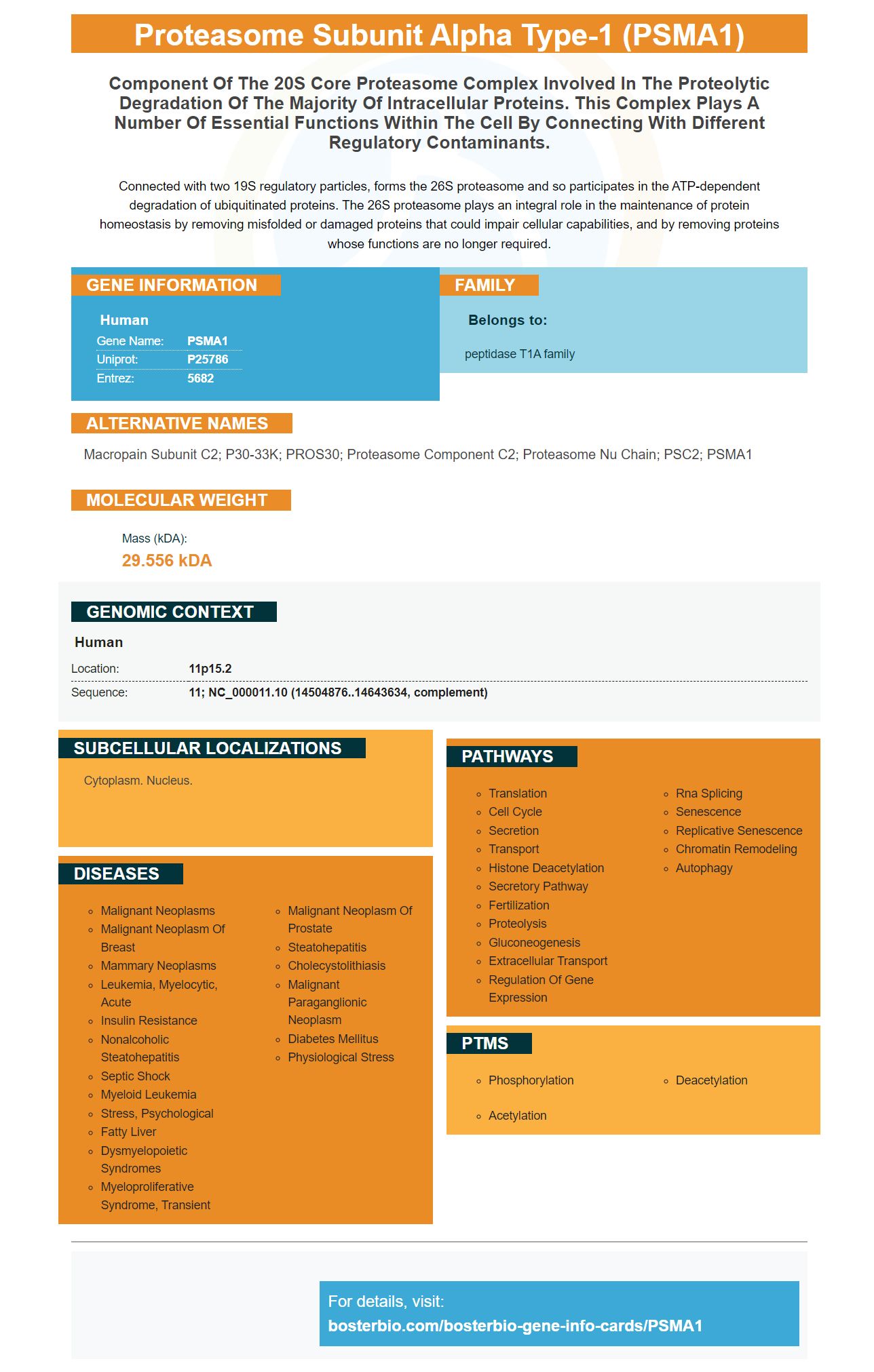
Facts about Proteasome subunit alpha type-1.
Connected with two 19S regulatory particles, forms the 26S proteasome and so participates in the ATP-dependent degradation of ubiquitinated proteins. The 26S proteasome plays an integral role in the maintenance of protein homeostasis by removing misfolded or damaged proteins that could impair cellular capabilities, and by removing proteins whose functions are no longer required.
Human | |
---|---|
Gene Name: | PSMA1 |
Uniprot: | P25786 |
Entrez: | 5682 |
Belongs to: |
---|
peptidase T1A family |
Macropain Subunit C2; P30-33K; PROS30; Proteasome Component C2; Proteasome nu Chain; PSC2; PSMA1
Mass (kDA):
29.556 kDA
Human | |
---|---|
Location: | 11p15.2 |
Sequence: | 11; NC_000011.10 (14504876..14643634, complement) |
Cytoplasm. Nucleus.
The human PSMA1 protein was expressed in E.coli with a His-Tag, a protein that can be used to determine the presence of the PSMA1 gene. For up to one week storage, keep at 2degC-8degC or -20degC-80degC long-term storage. Avoid repeated freeze-thaw cycles. The human PSMA1 protein is soluble in 20mM Tris-HCl buffer, containing 10% glycerol, 2mM DTT, and 0.15M NaCl. Scientists around the globe can use Boster product credits.
Researchers found that PSMA1 expression was significantly higher in eight of ten samples from colon tumor tissue. However, the protein was also found throughout normal mucosa. A Nature Communications study has shown that PSMA1 expression in cancer cells is 1.2-fold higher in normal colon tissue than in normal. These results suggest that PSMA1 could be a useful marker for colon cancer risk.
The study included two patient specimens, one male (and one female) with stage II or IV colon adenocarcinomas. The same markers were then applied to a larger group of colon cancer patients. These results suggest that PSMA1 expression may be an important biomarker for colon cancer. But, the research has not been conclusive. Further studies are needed in order to determine if PSMA1 has the ability to detect colon carcinomas more effectively.
PSMA1 was detected in the nuclei, or cells that are primary colon carcinoma cells. Normal mucosa did not have this expression. Its nuclear expression was stronger in tumor cells located near the invasive tumor front. In contrast, the protein was undetectable in mucosa from the normal colon. Normal mucosal tissues are normally negative or show weak to moderate PSMA1 activity.
The findings suggest that PSMA1 expression increases in colon cancer. The study uses matched pairs containing colon cancer tissues taken from patients who have had their colon removed. The patients with liver metastases also had cancer tissues that were isolated and subjected to immunohistochemistry. PSMA1 expression rises in colon cancer. This is a sign that the tumors have an immune reaction to the PSMA1 antibody.
Boster Bio published a study that found that PSMA1 was frequently mutated in metastatic cancers. These findings are consistent in previous research and suggest that PSMA1 might be a potential cancer marker. However, further studies need to be conducted in the presence of cancer to determine if PSMA1 expression increases. These findings are the first to show the utility of PSMA1 for colon cancer.
This study examined the differential expression of LAP3 within 8 colon cancer tissues. It also matched normal tissue. The expression level of LAP3 was found to be elevated in eight of the eight samples. The LAP3-positive cancer cells had an ER value of 6.6, which was higher than that of normal colon mucosa. LAP3 expression in cells was also significantly higher that normal mucosa.
The current study involved matching pairs of normal and cancerous colon tissue from patients who had undergone surgical colectomy. Patients were also provided with liver metastases. These tissue samples were processed for their protein extracts. Proteins were separated by 2D gel electrophoresis and assessed by 1D SDS-PAGE and immunohistochemistry. We used a cloned method for immunohistochemical confirmation.
Neoplastic colon cancer had a significantly higher level of maspin protein than normal mucosa. The tumor cells located at the invasive end of the tumor also showed an increased expression of Maspin. However, maspin expression is low or non-existent in healthy colonic and normal mucosa tissues.
These findings support the hypothesis that LAP3 is a tumor suppressor and can be a useful tool in the diagnosis and treatment of colon cancer. It is possible that LAP3 could be difficult to treat due to the absence of a specific marker. The Boster Bio Antibody to LAP3 is available for treatment of colon cancer and related diseases. This antibody was tested and validated in multiple platforms and has high affinity for human, mouse and rat cells.
The study involved serum from a stage 4 colon cancer patient being compared with sera taken from healthy individuals as well as a control group. After being blotted against cancer cells with autoantibodies, the samples showed dramatically different reactive patterns. These spots were largely made up of cancer-specific antibodies. The researchers concluded that this protein may be responsible for the increased expression of LAP3 in colon cancer.
Recently, it was discovered that ANXA3 levels are higher in colon cancer patients. These findings suggest that ANXA3 may be involved in the progression and development of colon cancer. High levels ANXA3 expression may be associated with colon cancer growth and metastasis. This protein may also be involved in the regulation and angiogenesis of tumours via HIF-a signalling pathway.
In addition, knockdown of ANXA3 knockdown inhibits the phosphorylation of NF-kB p65, an indicator of activation of the canonical NF-kB pathway. This could be why there is an increase in p65 and decreased IkBa. In this study, IkBa was knocked down in the MDA-MB-231 cell line using a specific shRNA lentivirus.
Interestingly, ANXA3 proteins are expressed in both normal tissue and cancerous tissue. Tumours tend to have the higher molecular weight protein. While most tumours express both forms of ANXA3, some cells only express one or the other. Le Cabec et al. Both forms of ANXA3 could be expressed in human leukemic myeloblast HL-60 cell lines.
Despite the fact ANXA3 has been implicated in the growth of colon cancer it is not known if it is a tumour suppressor. This cancer-suppressive drug Ox is used in the treatment of colorectal tumors. Moreover, the two CRC cell lines tested for resistance to Ox exhibited increased ANXA3 expression. These results suggest that ANXA3 might be an important drug target in the fight against cancer.
Interestingly, ANXA3 was also detected in gastric cancer. This gene is highly expressed in these tumours and is positively associated with tumour volume and TNM stage. Further studies are needed to determine the role ANXA3 has in gastric cancer. This study supports ANXA3 as an essential component of colon cancer progression.
These studies also showed that ANXA3 was associated with HCC multidrug resistance. The association between drug resistance and ANXA3 is being further investigated. These data offer new clues to the development of MDR in other cancers. Doctors should be aware of ANXA3 gene expression in colon carcinoma patients. Many other studies have also shown that colon cancer is linked to this cancer suppressor.
Cancer cells are able to switch off the angiogenic switch by activating IRE1dependent signaling pathways. Moreover, ER stress can cause a rapid expansion of regenerative intestinal-stem cells if IRE1a has been overexpressed. This phenomenon is independent of homeostasregulation. Thus, increased colon cancer cell proliferation can be attributed to ER stress-induced hyperactivation IRE1a.
PMID: 1888762 by DeMartino G.N., et al. The primary structures of four subunits of the human, high-molecular- weight proteinase, macropain (proteasome), are distinct but homologous.
PMID: 2025653 by Tamura T., et al. Molecular cloning and sequence analysis of cDNAs for five major subunits of human proteasomes (multi-catalytic proteinase complexes).