This website uses cookies to ensure you get the best experience on our website.
- Table of Contents
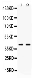
Facts about Phosphoserine aminotransferase.
Human | |
---|---|
Gene Name: | PSAT1 |
Uniprot: | Q9Y617 |
Entrez: | 29968 |
Belongs to: |
---|
class-V pyridoxal-phosphate-dependent aminotransferase family |
EC 2.6.1.52; EPIP; Phosphohydroxythreonine aminotransferase; phosphoserine aminotransferase 1; phosphoserine aminotransferase; PSAMGC1460; PSAT; PSAT1; PSATD; PSATendometrial progesterone-induced protein
Mass (kDA):
40.423 kDA
Human | |
---|---|
Location: | 9q21.2 |
Sequence: | 9; NC_000009.12 (78297125..78330093) |
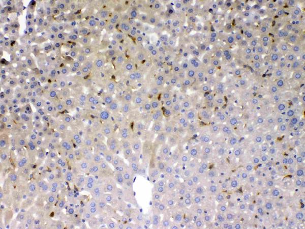
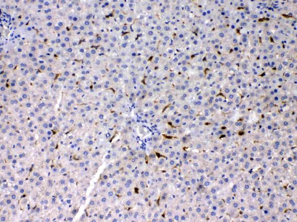
Expressed at high levels in the brain, liver, kidney and pancreas, and very weakly expressed in the thymus, prostate, testis and colon.
The PSAT1 gene encodes the PSAT1 marker. It is a class V pyridoxal-phosphate-dependent aminotransferase which plays a key function in the metabolism of glutamine. The enzyme is used in a variety of ways and is a prognostic marker for LGGs. We'll be discussing its top uses and ways to obtain it.
The PSAT1 gene is responsible for the creation of the enzyme Phosphoserine glucuronate, which is a major ingredient in the phosphate. Neu-Laxova syndrome 2, also known as NLS2, is a rare, fatal disorder caused by mutations in the genes PSAT1. NLS2 patients are also afflicted with ichthyosis, intra-uterine growth retardation, and leg deformities. PSAT1 mutations can cause congenital microcephaly and hypertonia, seizures, paramotor retardation, as well as abnormalities in the nerve system.
The brain, testis, prostate, thymus, and prostate all have the PSAT1 gene. Its expression is less prominent in the colon and prostate. Both kinds of PSAT1 expression are found in human cell lines, and they are linked with tumor regression. The gene PSAT1 is also involved in catabolism, a process that is known as serine biosynthesis. Antimetabolite purine antagonists (AMPs) are used to combat chemoresistance resulting from overexpression of PSAT1.
In humans, PSAT1 is a pyridoxal phosphate-dependent cytosolic protein that catalyzes the conversion of 3-phosphohydroxypyruvate into phosphoserine. This enzyme converts phosphoserine serine, which is the third step in the serine synthesis pathway that has been developed in the de novo stage. It forms an a/b homodimer with an catalytic-lysine as well as bound PLP to its active site. Humans express PSAT1 in liver and pancreas, kidneys and in the brain. The PSAT1 gene is affected by mutations that can cause the development of the neurodegenerative disease Neu-Laxova syndrome.
Humans have two orthologs: PSAT1 and PSAT2 and the gene PSAT1 codes for both. Both PSAT proteins can be found in the plastids. The PSAT1 protein can be found in tissues and cells. It is also found in keratinocytes and sperm cells.
This enzyme is also found in plants. PSAT1 and PSAT2 encode the PSAT gene. Humans have one PSAT1 gene, and two for PSAT2. The Arabidopsis PSAT1 has high homology to human PSAT1 and other species' PSAT genes. The Arabidopsis PSAT gene is also present, and the genome of this plant includes the peptide thought to be a target for localization into plastids.
PSAT1 is one of the human genes that produces a specific enzyme called Phosphate Kinase. PSAT1 is found in both the skin and muscles. It is involved in the metabolism of phosphate-glucosamine, one of the common building blocks for phosphate. It is also present in the brain.
Human liver produces PSAT1 phosphorus-kinase, which is a gene. This enzyme is essential in the phosphorylation of the phosphate. It is a major enzyme that plays a role in the transport of glycine and phosphate. The absence of it in the human body can be linked to various diseases.
The protein encoded by pyridoxal_deC is a nonribosomal peptide synthetase module. This protein also belongs to the decarboxylase family II. It shares homologues with glutamate decarboxylase and cysteine-sulfinic aciddecarboxylase. They all share similar catalytic residues.
The protein contains three natural organic compounds (pyridoxal phosphate, aldimine , and ketimine) that act as coenzymes in a variety of reactions. It plays a classical role in transamination and is involved in the metabolism of neurotransmitters. Its three-dimensional folds enable it to associate with other enzymes in an intricate manner.
The PLP cofactor can be attached to Lys219 via a Schiff base. The asn159 sidechain interacts with the nitrogen of the PLP rings, while the Asp187 residue interacts with the phenolic oxygen in Ala87. In addition the phosphate group of the PLP interacts with the nitrogen backbone of Ala87 and the side chains of Lys227, Ser88, and His27.
MycA is an evolutionary conserved area of a family enzymes that covalently attaches pyridoxal and lysine. While the mechanistic properties of the other pyridoxal-phosphate-dependent aminotransferases are similar however, each subfamily is different.
The active site of PLP is located at the interface between domains of the enzyme. Fold type I PLPs may be heterodimers or homodimers. The active site is found in the crevice that divides the two subunits. Aspartate aminotransferase was the very first aminotransferase that was PLP-dependent to be discovered.
Arginine oxidases belong to the protein family aspartate aminotransferase. The position C4 in PLP is the best for superoxide rebound. This is the ideal position for protonation. However other enzyme families may not exhibit isotopic enhancement. So, the protein family of arginine oxidases could evolve to utilize different substrates in the near future.
PacE and PacS produce an antibiotic known as peptidyl nucleoside pacidamycin. These enzymes are loaded with numerous other PLP dependent aminotransferases that could be used as targets for drugs. With the rise of antipsychotic drugs PLP-dependent aminotransferases have been targeted for their potential metabolic significance.
The Beta-elim family is distinguished by a significant remodeling of the protein's surface. They also offer a broad variety of possible interactions between proteins. Their models protein-like domains can be found at the C-terminal of multidomain modules. Phe112 in tyrosine-phenol-lyase is conserved, but does not contain the side chains that are required to catalyze.
Arginine oxidases are PLP-dependent enzymes that make use of molecular oxygen in catalysis. In addition, arginine oxidases are PLP-dependent and play a role in biosynthetic pathways. These enzymes can be broken into desaturases (hydroxylases) and oxidases. Among them, Ind4 is an exemplifying enzyme that oxidizes 4,5-dehydro-2-iminoarginine to imino-arginine.
The latest study of LGG patients suggests that overexpression of PSAT1 is a potential prognosis marker in certain cases. LGGs with IDh2 mutations and LGGs with 1p19q codeletion demonstrated significantly better outcomes compared with patients with wild-type LGGs. The findings were also confirmed by other studies in the same cohort. While the status of codeletion on chromosome 1p19q did not affect overall survival The TGGA data set included LGG patients with high levels of IDh2-mutated LGGs and those with no mutations.
The 2016 WHO classification of CNS cancers recommended that prognostic markers include IDh2 mutation status and chromosome 1p19q gene codeletion. This method may not be suitable for every case and has limitations. However, PSAT1 expression levels were significantly associated with a lower overall survival, and a higher five-year survival rate than PHGDH or PSPH.
The PSAT1 gene's overexpression is associated with changes to the IDh2 gene. While wild-type IDh2 converts isocitrate in a-KG and mutant IDh2 produces R-2HG. Overexpression of PSAT1 can result in an increase in R-2HG production and possibly resistance to chemotherapy and radiation.
IDh2 mutations in LGG patients improve their survival rate and response to therapy. They are found in 70 percent of LGGs and secondary Gliomas. There is no way to tell how these mutations can be a positive result. The findings are positive. They have shown that IDh2 mutations can be used as a prognostic marker for LGGs.
While there are many factors that predict LGG survival however, only the subset of B-cells was associated to less favorable outcomes. Other factors that were associated with lower outcomes in LGGs included age and the presence a large amount of lymphocytes with immune infiltration. PSAT1 is an independent marker for LGG prognosis.
The PSAT1 gene is controlled by the first two serine-synthetic proteins, PSPH, and a-KG. PSAT1 is a key factor in the growth and metastasis of HCC cells and provides potential therapeutic targets. PSPH is the second enzyme that regulates PSAT1 in the TCA cycle. PSAT1 has also been associated with a better outcome in LGG treatments.
PMID: 12633500 by Baek J.Y., et al. Characterization of human phosphoserine aminotransferase involved in the phosphorylated pathway of L-serine biosynthesis.
PMID: 17436247 by Hart C.E., et al. Phosphoserine aminotransferase deficiency: a novel disorder of the serine biosynthesis pathway.