This website uses cookies to ensure you get the best experience on our website.
- Table of Contents
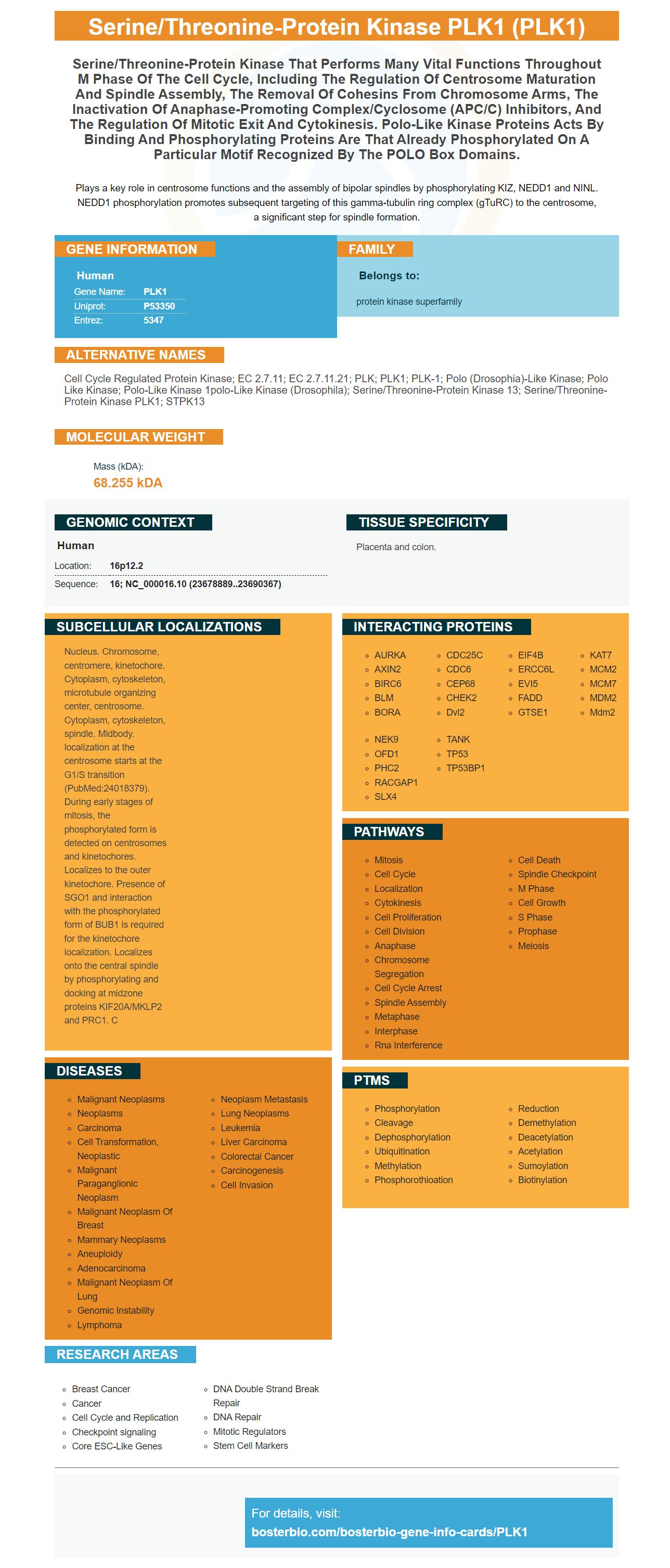
Facts about Serine/threonine-protein kinase PLK1.
Plays a key role in centrosome functions and the assembly of bipolar spindles by phosphorylating KIZ, NEDD1 and NINL. NEDD1 phosphorylation promotes subsequent targeting of this gamma-tubulin ring complex (gTuRC) to the centrosome, a significant step for spindle formation.
Human | |
---|---|
Gene Name: | PLK1 |
Uniprot: | P53350 |
Entrez: | 5347 |
Belongs to: |
---|
protein kinase superfamily |
cell cycle regulated protein kinase; EC 2.7.11; EC 2.7.11.21; PLK; PLK1; PLK-1; polo (Drosophia)-like kinase; polo like kinase; polo-like kinase 1polo-like kinase (Drosophila); Serine/threonine-protein kinase 13; serine/threonine-protein kinase PLK1; STPK13
Mass (kDA):
68.255 kDA
Human | |
---|---|
Location: | 16p12.2 |
Sequence: | 16; NC_000016.10 (23678889..23690367) |
Placenta and colon.
Nucleus. Chromosome, centromere, kinetochore. Cytoplasm, cytoskeleton, microtubule organizing center, centrosome. Cytoplasm, cytoskeleton, spindle. Midbody. localization at the centrosome starts at the G1/S transition (PubMed:24018379). During early stages of mitosis, the phosphorylated form is detected on centrosomes and kinetochores. Localizes to the outer kinetochore. Presence of SGO1 and interaction with the phosphorylated form of BUB1 is required for the kinetochore localization. Localizes onto the central spindle by phosphorylating and docking at midzone proteins KIF20A/MKLP2 and PRC1. C
This article will describe how Boster Bio's educational resources can help you use the PLK1 Marker for your research. These educational resources are accessible as PDFs and can be shared with others. Be sure to mention and hyperlink to Boster Bio when using its materials. The PLK1 gene is necessary for a variety of processes and functions. The PLK1 gene can be found in the human brain.
The Boster Bio Cell-Based ELISA test for PLK1 is a fast, efficient and reliable instrument for monitoring the PLK1 expression pattern. It measures the relative amount of PLK1 in cultured cells and can also be used to evaluate the effects of various treatments or activators. The kit includes a 96-well clear-bottom microplate for cell culture that contains 10x TBS, 1x Clear Quenching Buffer and 1x Clear Blocking Buffer.
The Boster Bio Cell-Based ELISA for PLK1 Marker employs antibodies that bind to RFP, an indicator of PLK1 activity. RFP is a frequent target for pharmaceutical trials because antibodies attach to it. The kit is sold at pharmaceutical companies like Merck, Sigma-Aldrich, and R&D Systems. Other brands include Flukasa and Sigma-Aldrich.
Boster Bio Cell-Based ELISA Kit for PLK1 Marker is compatible to different sources and compatible with various reagents. Among the most common PLK1 markers are CEP68, CDK1 phosphorylated Fry and CDK1-phosphorylated DCTN1. This kit also permits the determination of DNA-binding activities.
If you are in the market for an antibody against the PLK1 marker, you need to start by looking at the available antibodies. The PLK1 marker is a protein expressed in E. coli. If you know which proteins the PLK1 marker reacts to you can buy one of these antibodies. This antibody is specific for the human PLK1 protein. It can be purchased in whole cell lysates as well in suspensions and dilute solutions.
PLK1 is an anaphase-promoting (APC) Ubiquitin ligase. It also phosphorylates APC subunits Aurora A, cdc20, that are known to regulate the entry of mitotic cells. Plk1 has been shown to phosphorylate APC subunits during several studies, which could be a role for mitotic exit regulation. S-phase is stopped by the phosphorylated Ser137Asp mutation. The Thr210Asp mutation is resistant to DNA damage inhibition.
The development of a non-phosphorylatable anabolite peptide of the PLK1 marker has a number of advantages. It firstly, it avoids steps of electrophoresis or excision. This reagent can be used to capture of thiol-containing proteins on iodoacetylagarose beads. The peptide mixture is then subjected mass spectrometry. This reagent is extremely sensitive, allowing detection of more than 70 Cdk1 substrates.
The discovery of an as-allele unable to undergo phosphorylation might provide an excellent tool for localization studies. However, it has several disadvantages. For instance, homogenous substrate knockdown in human cells is a challenging process because of the difficulties in identifying specific phosphorylation sites in the cell of the target. It can also result in false-positive or false-negative results.
In humans Humans, an phosphorylatable peptide analog of the PLK1 marker is also in development that can detect pT182-Polo in all stages of the cell cycle. It is well-known that phosphorylation of Map205 by Cdk1 in embryonic cells and in the cultured cells is more pronounced.
The CNH domain of the PLK1 marker is undetermined. However, its structure suggests that it functions as the hub for multiple interactions between proteins. It is also believed to be involved with scaffolding proteins that belong to the WD40 family. Its presence in the structure of proteins is consistent and has shown results of binding. Therefore the team of researchers hopes to develop an PLK1 inhibitor based on this new inhibitor.
Polo-like kinases (PLKs) are a class of serine/threonine proteins kinases that are important regulators of cell cycle progression, mitosis, cytokinesis, and DNA damage response. They are widely expressed and found in a variety of tissues. It is therefore an important research area for therapeutic research.
The compounds are composed of two domains joined by an intermediary. The two domains compete for binding sites with one another and produce bifunctional inhibitors. Their high affinity for ATP is coupled with their specificity for the substrate. As a result, bifunctional inhibitors have to be highly selective to distinguish from proteins that are closely related. The length of the linkage, that connects the two domains can also affect the inhibitor's effectiveness.
BI 727 is a dihydropteridinone that interacts with PLK1 during mitosis. It binds to Plk1's hinge region by forming two hydrogen bonds on Cys133. The interaction results in catalytic inactivation of Plk1. The resulting phosphorylation inhibits the cell cycle and leads to an apoptosis. The compound has produced promising results in preclinical trials for patients suffering from advanced solid tumors.
As a specific small molecule for cancer drug, PLK1 has tremendous potential. It plays an essential role in cell division, and is an attractive target for targeted therapies. Bioactive domains in it regulate ATP binding and enzyme activation. The PLK1 marker is an excellent option for therapeutic development due to its crucial role in cell division. Why is it crucial to develop inhibitors for the PLK1 marker?
The PLK1 gene is a highly conserved genetic location that can be detected by various t andh2> dic located on both chromosomes in eukaryotes as well as humans. Although there are several genetic mutations that are associated with PLK1, many of them haven't been discovered. However, researchers have discovered that the PLK1 gene could be present on many chromosomes.
PLK1 activity is increased in cells as a result of the remodeling of the pericentriolar tissue at the mother spindle pole. As this protein is highly expressed in the body, the increased activity in this region could lead to cell-cycle arrest or premature aging, and even cancer. Despite the complexity there is a chance for its discovery. The PLK1 marker has numerous therapeutic possibilities.
PLK1 is elevated in mammalian cells during mitosis. PLK1 accumulates in the spindle poles and the kinetochores in metaphase, and transitions to the cytokinetic furrow after exiting metaphase. The PLK1 gene is also present in the midbody region, which is associated with the formation of the midbodyh2>n vitro studies have revealed that PLK1 is located mostly at the spindle poles during mitosis. However, this does not always correspond with what happens in living.
Numerous biological assays have been developed to detect PLK1 in various animal samples. Boster Bio has developed antibodies against the PLK1 marker using rabbit and mouse as model animals. These antibodies react with PLK1 in a variety of biological assays. Here are a few of the most useful applications of the PLK1 marker. These include: (i) Detecting the presence of PLK1 in plasma and human serum; (i) Measurement of its presence in brain tumors and other cancers;
Peptide inhibitors are the first class of biological chemicals that block Plks. They are able to bind to Plk1 PBDs with high affinity, and inhibit the activity of the Plk1 molecule. Several inhibitors have been discovered by using this technique, including synthetic poloxin and Thymoquinone. The PLK1 substrate binding site can be targeted by inhibitors.
PMID: 8018557 by Hamanaka R., et al. Cloning and characterization of human and murine homologues of the Drosophila polo serine-threonine kinase.
PMID: 7902533 by Lake R.J., et al. Cell cycle- and terminal differentiation-associated regulation of the mouse mRNA encoding a conserved mitotic protein kinase.