This website uses cookies to ensure you get the best experience on our website.
- Table of Contents
8 Citations 8 Q&As
1 Citations 1 Q&As
1 Citations 6 Q&As
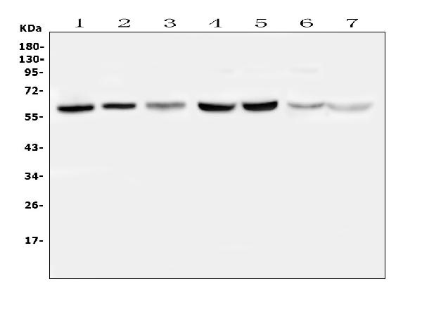
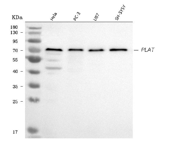
Facts about Tissue-type plasminogen activator.
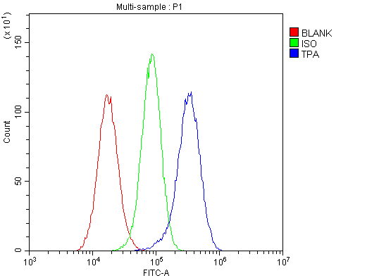
Plays a direct role in facilitating neuronal migration. .
Human | |
---|---|
Gene Name: | PLAT |
Uniprot: | P00750 |
Entrez: | 5327 |
Belongs to: |
---|
peptidase S1 family |
Alteplase; DKFZp686I03148; EC 3.4.21; EC 3.4.21.68; plasminogen activator, tissue type; plasminogen activator, tissue; PLAT; Reteplase; tissue plasminogen activator (t-PA); tissue-type plasminogen activator; TPA; T-PA; tPlasminogen Activator; t-Plasminogen Activator
Mass (kDA):
62.917 kDA
Human | |
---|---|
Location: | 8p11.21 |
Sequence: | 8; NC_000008.11 (42174718..42207565, complement) |
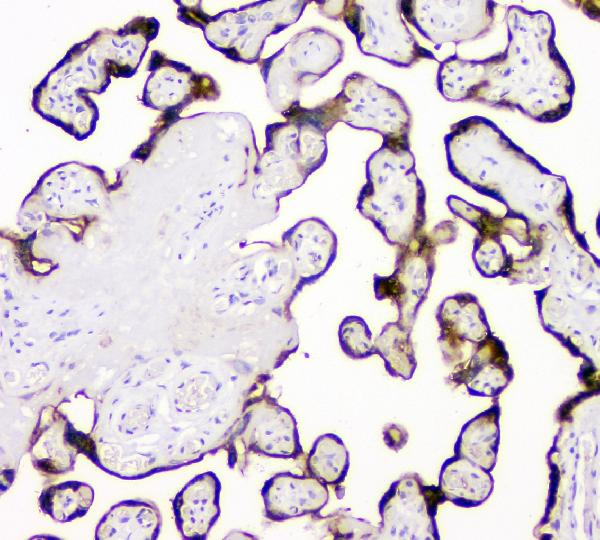
Synthesized in numerous tissues (including tumors) and secreted into most extracellular body fluids, such as plasma, uterine fluid, saliva, gingival crevicular fluid, tears, seminal fluid, and milk.
Secreted, extracellular space.
Boster Bio ELISA and antibodies are one of the most popular diagnostic tools in today's labs. The company has been in business since 1993, producing quality antibodies and kits for ELISA that can be used in a variety of applications. Boster Bio is a manufacturer of antibodies and reagents that have been tested with rigorous methods and have been awarded over 29,000 citations. Alongside a wide array of applications, the Boster Bio kits can be beneficial in cancer research.
Although it's easy to use biotinylated-lectins to detect samples, there are dangers. Unfortunately, these methods could result in disappointing results. Many researchers have stated that they haven't been able to achieve the desired results in as many as 25% of their cases. Bioster Bio has come up with a variety of ways to improve the efficiency of protein transfer using membrane staining.
The first method involves staining the membranes using a solution that contains 0.2% Ponceau S and 5% glacial acetic acid. The membranes are then placed in a scanner for determining the optical density of the stained proteins. The results of two tests were compared to verify that the membranes are able to bind the target protein in the absence of any other proteins. Afterwards, the re-probing was carried out to determine if the staining method on membranes improved the protein transfer efficiency.
In the third method Bioster has increased protein transfer efficiency by testing the ability of membranes to attract specific antibodies. They have demonstrated that the PVDF membrane is more effective than the NC membrane in the detection of multiple targets. The PVDF membrane also had a higher reprobing capability than the NC membrane. These results suggest that the PVDF membrane has a higher re-probing ability than the NC membrane, while the NC membrane was more sensitive in three other instances.
The next step to increase efficiency of transfer of proteins through membrane staining is to conduct an electrophoretic titration. A buffer containing methanol can be used to conduct electrophoresis. This buffer is used to titrate proteins, and facilitate their transfer to the membrane. The PVDF membrane assists in the formation of conjugates of negatively and positively charged proteins, while the nitrocellulose membrane is free of negative staining.
Boster Bio's ECL Chemistry chemiluminescent detection device is used to detect proteins in biological samples. The ECL Chemistry combines primary antibodies that recognize a protein with secondary antibodies that are labeled with HRP or luminol-based HRP. The primary antibodies are incubated with the ECL Detection Reagents and result in the formation of a luminescent light that could be captured on CCD cameras or x-ray film.
The X-ray film is a popular chemiluminescent substrate used in HRP, a technique that is based on two components that include a stable solution of peroxide and an enhanced substrate solution, often a luminol-based product. The working solution is created by mixing equal volumes of each component. The reaction produces light with a wavelength of 425nm. The ECL system can also be captured using CCDs that have been cooled as well as X-ray imaging cameras. These instruments can capture large quantities with ease and speed which is a significant advantage over film.
The Clarity Max are compatible with horseradish peroxidase conjugates. These chemistry systems can be utilized for any type of western blot. Advanced chemiluminescent detection systems depend on the emission of light to indicate the presence of protein in an extract. They are a standard tool used for protein detection on western blots. The second stage of the reaction involves combining the secondary antibody to the horseradish peroxidase enzyme. The horseradish peroxidase is able to react with the substrate, resulting in the formation of a signal at 428 nanometers. This signal is then detected on a digital imager , or X-ray film.
The Boster Bio's ECL chemistry detects protein-derived molecules with a single color-based reagent. The ECL reagent reacts with the target protein by the reaction of HRP with the protein substrate. This light emission occurs when the primary antibody binds to the protein target. This reagent is chemiluminescent and allows for multiple exposures, which increases the signal to noise ratio. Chemiluminescent Chemistry has the highest sensitivity potential, in comparison to other chemical detection methods. The advantages make it the detector preferred by many research laboratories for protein.
Boster antibodies offer several advantages over other chemical reagents. They can first increase the intensity of the signals produced by an enzyme. They can also increase the sensitiveness of the reaction, leading to higher signal intensity and greater the sensitivity. The Boster ECL chemical reagent suitable for western blots where the proteins of interest are abundant.
Boster's DAB Chromogenic detector system is used to stain tissue sections and cells for immunocytochemistry and applications using immunohistochemistry. This system has a stability rating of one year at temperatures of -20°C. Boster guarantees that the product performs as intended in the tested applications. You can also try it yourself by purchasing the sample kit. Learn more about the Boster DAB chromogenic detection system!
To use this chromogenic detection device, serum samples are reduced to 1:20 in 0.01 M PBS. Then, they are incubated at 37 degrees Celsius for one hour. The TMB chromogenic Solution then added to the sample. It is incubated at 37°C for 20 minutes. Then, the reaction is stopped by adding 2 M H2SO4.
To use the DAB chromogenic detection system, you must first prepare your samples. The sample should contain the antigen or antibodies you are trying to identify. To do this, use an antibody labeled with an enzyme. The substrate reacts with the enzyme and creates an insoluble colored substance that can be observed with a light microscope or electron microscope. The enzyme-antibody complex is the most important component of the chromogenic detection system.
PMID: 6337343 by Pennica D., et al. Cloning and expression of human tissue-type plasminogen activator cDNA in E. coli.
PMID: 6089198 by Ny T., et al. The structure of the human tissue-type plasminogen activator gene: correlation of intron and exon structures to functional and structural domains.
*More publications can be found for each product on its corresponding product page