This website uses cookies to ensure you get the best experience on our website.
- Table of Contents
5 Q&As
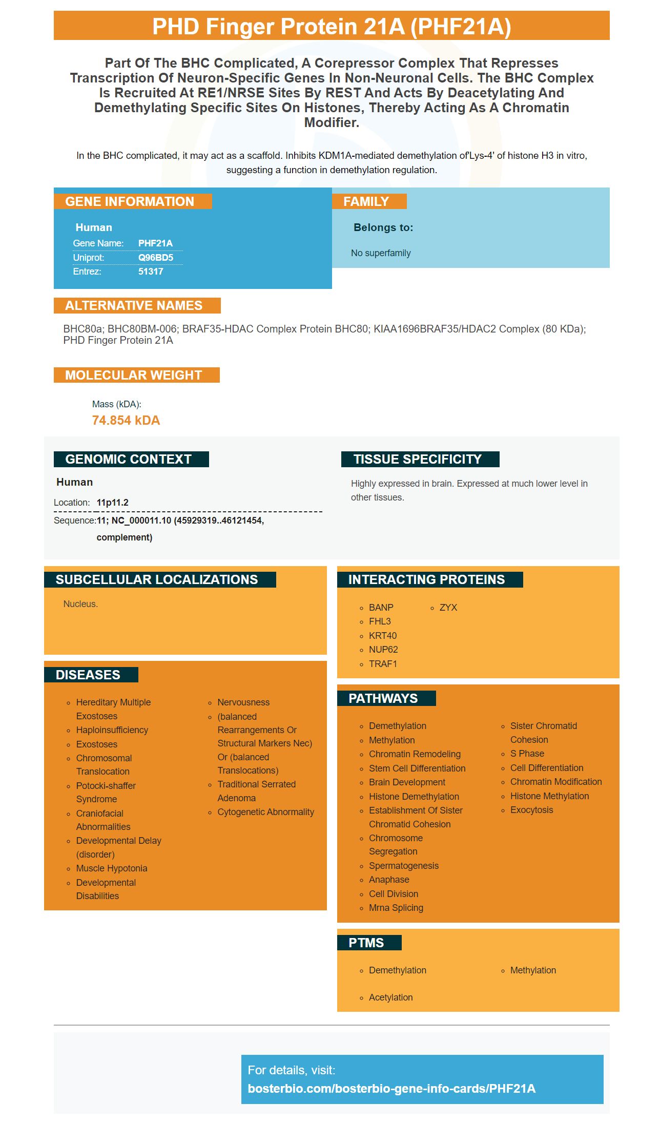
Facts about PHD finger protein 21A.
In the BHC complicated, it may act as a scaffold. Inhibits KDM1A-mediated demethylation of'Lys-4' of histone H3 in vitro, suggesting a function in demethylation regulation.
Human | |
---|---|
Gene Name: | PHF21A |
Uniprot: | Q96BD5 |
Entrez: | 51317 |
Belongs to: |
---|
No superfamily |
BHC80a; BHC80BM-006; BRAF35-HDAC complex protein BHC80; KIAA1696BRAF35/HDAC2 complex (80 kDa); PHD finger protein 21A
Mass (kDA):
74.854 kDA
Human | |
---|---|
Location: | 11p11.2 |
Sequence: | 11; NC_000011.10 (45929319..46121454, complement) |
Highly expressed in brain. Expressed at much lower level in other tissues.
Nucleus.
This article discusses the use Boster's high affinity primary antibodies to detect PHF21A. Learn how to clean and record your results on autoradiography film. This information is beneficial for both novice as well as experienced researchers. Boster's antibodies are often cited and validated in the research community. Read on to learn about the benefits of Boster antibodies for PHF21A ligand detection.
Traditional mIHC relies heavily on different species and isotypes of antibodies. Primary antibodies raised in different species of animals are used. Each marker also requires a different chromogen. Therefore, fluorescent mIHC only supports three markers in the filter sets. Fluorescent MIHC provides high content data for a single tissue section.
The primary antibody detects the target protein, while the secondary antibody amplifies the signal. The choice between primary and secondary antibodies is dependent on many parameters. For antibodies against Strep–tagged proteins, murine StrepMAB Classicic makes a great choice. It is not always the best option. Sometimes, a specific reagent is required to target a given protein.
Primary antibodies are immunoglobulins which bind to specific antigens. It is important to measure antibody quality by determining their affinity and specificity. Higher affinity signifies a better ability of the antibody to detect and measure antigens of interest. Poor specificity can mean that the antibody is unable to bind to unintended substances. A good primary antibody is capable of detecting, measuring, and purifying the targeted antigen.
Nonspecific hooking is a problem that can limit the sensitivity of an ELISA. High-affinity primary antibodies with PHF21A marker are not cross-reactive. The assay's sensitivity will also be lower. This is why background signals should be avoided when using this method. Dual-antibody detection has this advantage.
Three methods were used to determine the subcellular location of PHF21A. To identify variants, the diagnostic trio whole exome and Sanger sequencing was used. The AT Hook domain of the PHF21A genome was also identified. There was a low degree variation between these three methods. The PHF21A mark was found in the brain of an adult mouse and in the intestine. It was also expressed by multiple tissues.
The PHF21A protein is highly expressed within the fetal and perinatal brains. It is also found in lymphocytes and other parts. It is associated a range neurological phenotypes such as the cerebellum, occipital and cerebellum. PotockiShaffer syndrome has also been linked with the PHF21A mutation. This is a rare disorder that causes intellectual disability and unusual facial features.
The detection of the PHF21A genes has many uses. The markers are used to detect craniofacial disorders, including asymmetrical ear lobes. The gene is associated with craniofacial defects in two PSS subjects. It has also been associated with craniofacial ID and craniofacial irregularities. Different genetic tests have been developed that can detect the presence or absence of the PHF21A genes.
Whole exome sequencing was performed in four patients. After this, the PHF21A genes were detected by Sanger sequencing. After this, genomic DNA was fragmented and ligated to Illumina multiplex PE adapters. The pre-capture library was enriched using VCRome2.1 insolution probes, which were biotin-labeled. After the library had been created, the samples of the library were analyzed to confirm the presence or absence of the PHF21A marker.
Autoradiography uses a chemical substance, known as a Radioactive Tracer, to take a photograph of a radioactive resource within a sample. This requires close contact between your sample and the medium. Autoradiography relied in the past on film emulsions, mostly silver halide film. The film thickness, size of the silver-halide grains and performance of an optical densitometer all influence the resolution of autoradiography. Autoradiography film resolution is generally around 10 mm. Phosphor imaging, however, is a very efficient and convenient way to record results.
Autoradiography has been extensively utilized in many fields of medicine, biology, and medicine since its discovery. Cell biologists have used autoradiography to monitor DNA synthesis during the cell cycle. Researchers studying steroid hormones have used autoradiography to study how their effects affect the genetic machinery of target cells. Autoradiography has been used to discover the neuronal communication pathways by neurologists. This technology has been in radiology for more then 50 years.
An excellent way of recording medical images is to use autoradiography film. It is composed of an emulsion consisting of radiation-sensitive, transparent crystals. The film is then exposed for radiation to activate the material and create an imaging image. While radiation exposure doesn't change the film's image, it transforms its latent image to a visible image. The resulting image is saved as an electronic medical record.
Mass spectrometric imaging (MSI) is a relatively new method that is gaining popularity in the laboratory. Both techniques use whole-body tissues and both require dosed animals. MALDI-MSI does not require radioactive compounds. MALDI requires a matrix, which is not required for WBA. Although autoradiographers are unable to identify the parent molecule from the label, it is much more efficient than the former.
PMID: 12032298 by Hakimi M.-A., et al. A core-BRAF35 complex containing histone deacetylase mediates repression of neuronal-specific genes.
PMID: 12493763 by Hakimi M.-A., et al. A candidate X-linked mental retardation gene is a component of a new family of histone deacetylase-containing complexes.