This website uses cookies to ensure you get the best experience on our website.
- Table of Contents
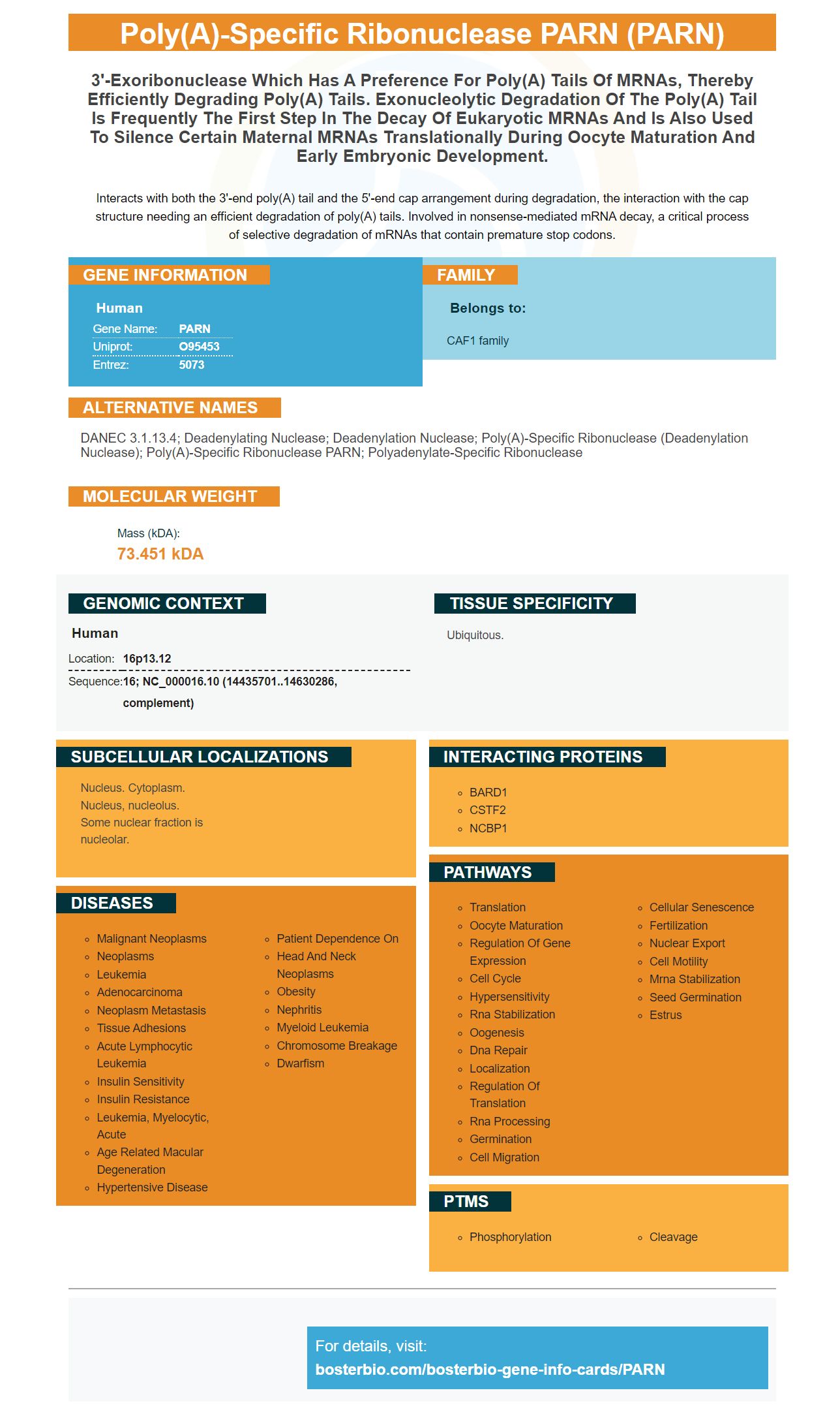
Facts about Poly(A)-specific ribonuclease PARN.
Interacts with both the 3'-end poly(A) tail and the 5'-end cap arrangement during degradation, the interaction with the cap structure needing an efficient degradation of poly(A) tails. Involved in nonsense-mediated mRNA decay, a critical process of selective degradation of mRNAs that contain premature stop codons.
Human | |
---|---|
Gene Name: | PARN |
Uniprot: | O95453 |
Entrez: | 5073 |
Belongs to: |
---|
CAF1 family |
DANEC 3.1.13.4; Deadenylating nuclease; Deadenylation nuclease; poly(A)-specific ribonuclease (deadenylation nuclease); poly(A)-specific ribonuclease PARN; Polyadenylate-specific ribonuclease
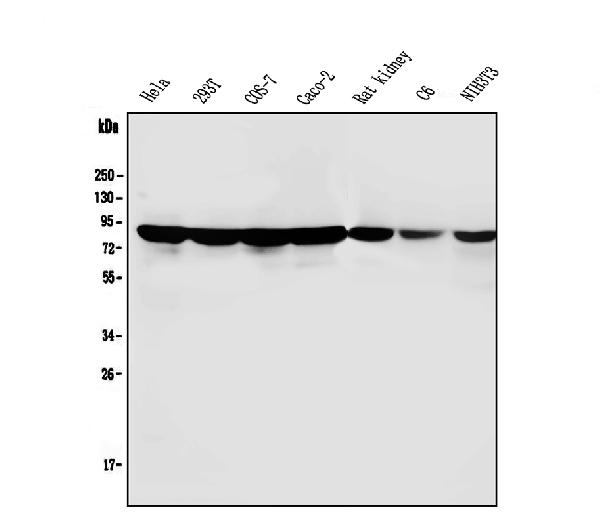
Mass (kDA):
73.451 kDA
Human | |
---|---|
Location: | 16p13.12 |
Sequence: | 16; NC_000016.10 (14435701..14630286, complement) |
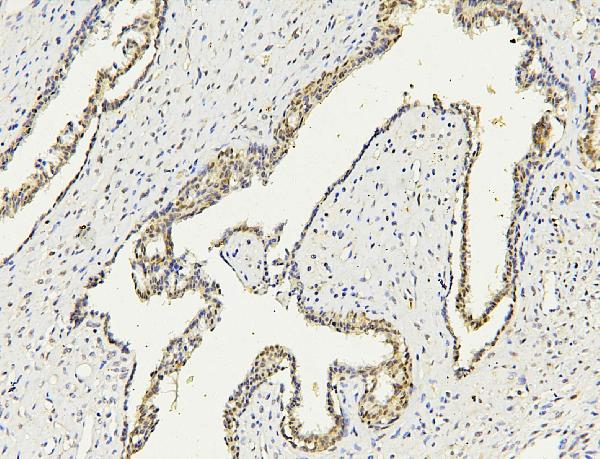
Ubiquitous.
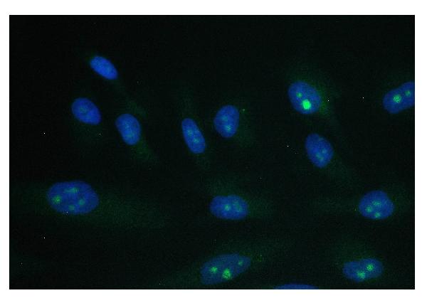
Nucleus. Cytoplasm. Nucleus, nucleolus. Some nuclear fraction is nucleolar.
PARN is now detectable by using biological assays. These assays use monoclonal or polyclonal antibodies that react to PARN in a variety of animal samples. The boster bio lab uses rabbit and mouse as the host species for their development of PARN antibodies. PARN is a product of exonucleolytic depoly(A) tails. This is a well-known way of blocking early maternal mRNA translation.
The CI MPR, also known as the cation-independent mannose 6-phosphate receptor, is a multi-functional protein that plays an important role in the sorting of lysosomal enzymes. The phosphomannosyl recognition system is crucial for the sorting process, and the discovery of the CIMPR has raised the possibility that it played an additional function. The CIMPR is similar to the insulin-like growth factor type-2 receptor and is also involved in the transportation of newly synthesized acid hydrolases to the prelysosomal compartment. When the ligand gets dissociated at low pH, the acid hydrolases that have been synthesized are packed into the lysosome.
The reactivity of this antibody was confirmed using pre-treatment with an synthetic ligand (a protin molecule) in the human cell extract. The reaction mixture was incubated at 4°C for at least 20 hours, with alternating the lysate-based washings. Western blot analysis of Hep-2 cell-derived lysates revealed no immunoprecipitated band.
In addition to its function in sorting acid hydrolases, the cation-independent mannose 6-phosphate receptor is also a key factor in the release of specific cellular proteins. The biosynthetic process of sorting and sorting enzymes that are phosphorylated can only be achieved by CI-Man6PR which is the sole receptor on cells.
The CI-Man6PR is located in a multi-peak distribution within the Percoll fraction. It is found primarily in early endosomes , as well as Golgi structures. However, smaller amounts are present in fractions five and 10. The fractions five and ten have lateral endosomes as well as prelysosomes.
The 78-kDa CIMan6PR protein was used to identify this protein in AD patients. This receptor is also referred to as LAMP2 and LIMPII. Particularly, this antibody targets the 78-kDa receptor. The anti-CI-Man6PR reactivity in the mouse was then identified and then analyzed using the analysis of immunoblots.
The CI Man6PR can also target acid hydrolases on lysosomes as well in addition to the Man6P-specific receptor that is dependent on cation. These enzymes can be directed to the plasma membrane by means of an 78-kDa receptor. This method is suitable for a variety of applications in research.
The CI-MPR/IGF-2R autoantibody is thought to be an autoantibody that causes an autoimmune response to exogenous Ag. The CI MPR/IGF-2R is a Th2-specific surface protein that regulates macrophage activation. It also blocks Th2-mediated auto or alloimmune response.
In addition, the CIMan6PR is highly stable. Numerous studies have demonstrated its stability. Anti-Mannose 6 Phosphate receptor (CI-Man6PR) in Boster Bio is compatible with the HEK-APPSwe cell line. This product can also be used for use in biochemical research. Its CI Man6PR is capable of recognising proteins of 78 kDa.
The CI Man6PR is located in the Golgi apparatus. The LIMPII is found in vesicles that surround the nucleus. Moreover, this molecule is closely connected to clusterin and HSPA1A which negatively influence APP processing. Clusterin and LIMPII also regulate APMAP production and processing.
IGF2R is a protein-coding gene which is essential in a myriad of diseases like Hepatocellular Carcinoma or Mucolipidosis Ii Alpha/Beta. It also has connections to G protein-coupled receptor activity, as well as to the CREB Pathway. Insulin receptors consist of a transmembrane beta subunit with intracellular tyrosine-kinase function. The insulin receptor forms a homodimer after insulin is applied to cells and signals the presence of IGF2R.
Multiple functions play CD86 in the immune system. It is necessary for the production and activation of cell-killer IL-2. It also stimulates T cell expansion. In addition, CD86 serves as a crucial costimulatory signal to determine the participation of T cells in immune responses. It also plays an important role in the reactivity of the immune system by modulating the expression of other markers. This article explores the use of anti-CD86 antibodies in the study of this receptor in the cellular.
CD86 is involved in the regulation of tolerance induction and T cell differentiation. In the murine SS model, CD86 stimulation inhibits the expression of PARN markers and increases the production of IL-4. Anti-CD86 treatment also inhibits autoimmunity-mediated Th2 progression by increasing the production of IL-4. The treatment for anti-CD86 does not affect the conventional IAV-specific T cell responses.
Anti-CD86 antibodies target murine CD86 which is a membrane protein produced by antigen-presenting cell. CD86 interacts with the CD28 antigen, and acts as a costimulatory molecule. However, lack of sufficient co-stimulation between anti-CD86 and antigen could result in tolerance. In addition to the immune response, CD86 can lead to diseases such as myocarditis and gallbladder cancer.
It isn't clear what the role CD86 plays in the proliferation of T-cells. CD86 has been found to block conventional CD4+FoxP3T T cells. The absence of CTLA-4 may be the reason for this. Interestingly, CD86 and CTLA-4 co-localize and this could be an important factor. It is still necessary to determine if anti-CD86 can be used to treat cancerous diseases.
CD86 is used in many areas of cancer research. For instance, it's used as a marker for the Treg subpopulation. It also acts as a costimulatory stimulant ligand to CD28-expressing T cells. This mechanism could also affect the survival and growth of T cells, specifically in patients suffering from inflammatory disease. Further research is required to understand the role of CD86 in the field of T-cell immunology.
Anti-CD86 moAbs can be used to monitor the process of disease and to prevent tumorigenesis. In one study, anti-CD86 MoAbs block the growth of inflammatory lesions in mice. These lesions were more pronounced in 3d-Tx mice than in control mice. This suggests that anti-CD86 antibodies play significant roles in the field of cancer immunotherapy. But it's still unclear which moAbs are effective in treating cancer and other inflammatory diseases.
Moreover, anti-CD86 treatment inhibits the growth of T cells that express the autoantigen recombinant a-fodrin. Anti-CD80 MoAbs however, on the other hand, had no effect upon the development of lesions. Anti-CD86 antibodies actually hindered the development of autoimmune exocrinopathy (NFS/sld) in mice. These findings are promising.
PMID: 9736620 by Koerner C.G., et al. The deadenylating nuclease (DAN) is involved in poly(A) tail removal during the meiotic maturation of Xenopus oocytes.
PMID: 10640832 by Buiting K., et al. The human gene for the poly(A)-specific ribonuclease (PARN) maps to 16p13 and has a truncated copy in the Prader-Willi/Angelman syndrome region on 15q11-->q13.