This website uses cookies to ensure you get the best experience on our website.
- Table of Contents
1 Citations
1 Citations
Facts about P2X purinoceptor 7.
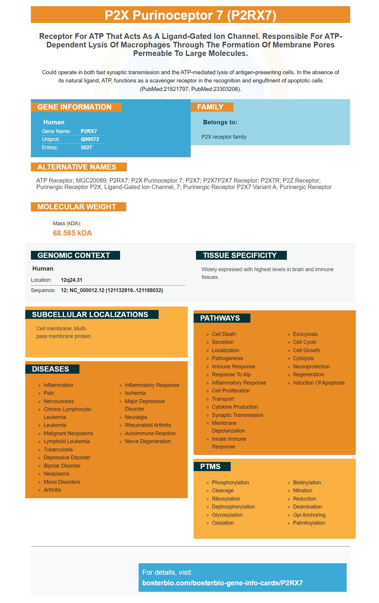
Could operate in both fast synaptic transmission and the ATP-mediated lysis of antigen-presenting cells. In the absence of its natural ligand, ATP, functions as a scavenger receptor in the recognition and engulfment of apoptotic cells (PubMed:21821797, PubMed:23303206).
Human | |
---|---|
Gene Name: | P2RX7 |
Uniprot: | Q99572 |
Entrez: | 5027 |
Belongs to: |
---|
P2X receptor family |
ATP receptor; MGC20089; P2RX7; P2X purinoceptor 7; P2X7; P2X7P2X7 receptor; P2X7R; P2Z receptor; purinergic receptor P2X, ligand-gated ion channel, 7; purinergic receptor P2X7 variant A; Purinergic receptor
Mass (kDA):
68.585 kDA
Human | |
---|---|
Location: | 12q24.31 |
Sequence: | 12; NC_000012.12 (121132819..121188032) |
Widely expressed with highest levels in brain and immune tissues.
Cell membrane; Multi-pass membrane protein.
If you've ever wondered about the best uses for the P2RX7 Marker, you're not alone. This article will introduce you to this ligand-gated ion channel, a scavenger receptor that functions as an important factor in the regulation of lymphocyte numbers and subtypes. You'll also learn that it's an excellent marker for astroglia in the brain.
The P2X7 ligand-gated ion channels have two distinct modes of function. In the absence of ATP, they function as scavenger receptors. As a ligand-gated ion channel, it is comprised of a trimer of subunits with two transmembrane domains.
These ion channels are activated by extracellular ATP. The release of this ATP signals the immune system to respond to tissue damage. This channel also promotes the survival and metabolic fitness of memory CD8+ T cells. When activated by ATP, P2X7 enhances cell-to-cell communication. As a result, P2X7 is involved in the regulation of cell growth and inflammation.
The purinergic receptor P2RX7 is expressed on glial cells and myeloid cells in the CNS. This receptor is implicated in autoimmune disease, as it promotes the activation of both glial cells and T-cells. This protein is expressed on immune cells, and when extracellular ATP reaches the brain, it acts on purinoceptors on neurons. The P2X7 ligand-gated ion channel is activated by high concentrations of ATP. The result of prolonged activation is the formation of a large cytolytic pore.
A new drug called emodin has inhibited the signaling activity of the P2X7 ion channel, and in this way, the treatment of HPDE6-C7 cells with emodin reduces the amount of the inflammatory proteins. It also inhibits the production of NLRP3 and ASC. This treatment also inhibits the production of ATP and reduces the toxicity of the drug.
The P2RX7 antibody is made from a synthetic peptide that is derived from a sequence of the rat P2RX7 protein. This sequence differs from the related human and mouse proteins by one amino acid. The resulting protein is expressed in the cell as P2X7, NLRP3 and caspase-1 by western blot.
The P2RX7 receptor plays an important role in phagocytosis. It has been shown that P2X7 receptors are essential for this function and are highly expressed in the surface of cells. Furthermore, the P2X7 receptor is required for CNS development in early pregnancy. Although P2X7 is an essential scavenger receptor, other scavenger receptors likely participate in CNS development and compensate for the genetic ablation of P2X7 in mice.
It is also important to note that the P2RX7R is a therapeutic target for HHV-6A infection. Moreover, P2X7R has been linked with infertility. In order to identify P2X7R as a potential therapeutic target, Boster Bio's P2RX7 marker functions as a scavenger receptor.
A meta-analysis of gene expression data of TCGA-GBM samples showed that P2RX7R is highly expressed in malignant tumors. In addition, only 5% of biopsies analyzed had mutations in this gene. However, RNA seq data showed that P2X7R was upregulated in both GBM and glioma. However, there were several associations between non-synonymous SNPs and a number of disease conditions, including hematopoietic tumors. As a result, consensus was reached that most malignant tumors overexpress P2X7R.
P2RX7 receptor is present in the membranes of intracellular organelles. This receptor has a known role in phagosome-lysosome fusion. Other organelle-specific functions may also be supported by the P2X7 receptor. The ATP-dependent cellular environment is likely to support P2X7-mediated phagocytosis.
The P2RX7 protein is a critical regulator of lymphocyte numbers and subtypes. Lack of this protein may promote the development of EAE, an autoimmune disorder characterized by an increase in lymphocyte numbers. In a previous study, P2x7 deficiency resulted in an increased frequency of disease. In the current study, researchers found that mice with an alternate form of P2X7 do not exhibit increased cytokine production in response to BzATP or other agonists.
Activated T cells infect the CNS with autoimmune demyelinating disease. As a result, they produce pro-inflammatory mediators, activate parenchymal glial cells, and cause neuronal and oligodendrocyte damage. The results also suggested that activation of the P2X7 receptor may be implicated in the development of multiple sclerosis (MS). In mice with P2x7 deficiency, the disease was induced in wildtype and P2x7-deficient mice. Clinical signs were observed and the development of disease progression was monitored by immunocytochemical staining of brain tissue and Tcell cytokine production.
As a result of the P2RX7-DNC, it has been shown that mutation of the first two cysteine residues renders the full-length pdP2X7 inactive in the presence of CTD. This demonstrates that palmitoylation is a possible mechanism for the facilitation of P2X7 channel activity.
In addition to regulating the numbers of lymphocytes, this marker also regulates the number of different types of inflammatory mediators. As a result, the P2RX7 gene has a significant impact on the number of immune cells, as demonstrated by the P2X7 null mouse model. In addition, this gene is essential in regulating T-cell activation and the development of multiple sclerosis, a disease that is often characterized by autoimmune diseases.
Recently, scientists have discovered that the protein P2RX7 is expressed in the brain. The expression levels of this protein are regulated by glial cells, including astrocytes. This protein can be detected by real-time RT-qPCR, and its relative expression was measured by comparing it to the levels of endogenous murine P2RX7 mRNA.
The human P2RX7 receptor is composed of 10 different isoforms. Human P2RX7H is a non-neuronal protein and contains an exon 3 that is missing in other forms. The human P2RX7H presents a copy of exon 3 near the amino-terminal region. Hence, human P2RX7 is a useful marker of astroglia in the brain.
The human P2RX7 receptor has a unique phenotypic localization and participates in hypoxic/hypoglycemic processes. Its activity may be a sensor for dysregulated neuronal activity and ATP release during oxygen/glucose deprivation. Furthermore, P2RX7R is expressed in neurons and astrocytes.
Although the human P2RX7 gene has been linked to an increased risk of BD in some studies, it has been difficult to establish a definitive association between BD and the presence of P2RX7. Previous studies in the UK and Canada have shown that P2RX7 promotes gain of function. Furthermore, the polymorphism of rs2230912 is associated with the symptomatic stage of BD patients.
Recent studies have shown that P2RX7 is expressed in reactive microglia and may play a role in the upregulation of nitric oxide synthase and IL-1b. These studies indicate that ablation of the P2X7 receptor in mice can have differing outcomes in MS patients. A similar study in rats also showed that BBG reduced the inflammation-inducing cytokine release in patients treated with 50 mg of BBG.
Molecular research has identified a new therapeutic target for preventing HHV-6A infection. The virus replicates mainly in salivary glands and is latent in peripheral blood mononuclear cells. While laboratory studies for diagnosis of HHV-6 are unnecessary for healthy individuals, they are often necessary for immunocompromised individuals. Molecular tests for HHV-6A infection can be done by using GS cells, MOLT-3 cells, or HSB-2.
Recent studies have revealed that HHV-6A has a novel IE2 protein with a dimeric carboxyl-terminal domain that is similar to the DNA-binding domains of EBV EBNA1 and KHSV LANA. However, it is not known whether the HHV-6A IE2 protein acts similarly to the EBV EBNA1 and KHSV LANA proteins, which play similar roles during the replication of latent viral DNA.
Two L analogs of BDCRB and foscarnet were studied for their activity against HHV-6A and HHV-6B. While both CYP-derived RNA-binding proteins inhibit HHV-6A and B, they have lower toxicity in mice. In vitro data also revealed that cyclopropavir was superior to ganciclovir and cyprovir when compared to CDV control.
Primary HHV-6 infection is the most common cause of febrile illness among young children, and is associated with hospitalizations and febrile seizures. Most people are infected with HHV-6 during their lifetime, although there is no proven way to prevent infection. Nevertheless, reactivation of HHV-6A infection may occur at any age and is highly prevalent in different regions of the world.
Another promising therapeutic target for preventing HHV-6A infection is CMV-423. This non-nucleoside inhibitor is effective against HHV-6A and B and is in phase III clinical trials. This new therapeutic target is effective at subcytotoxic concentrations and has evidence for BBB penetration. The drug is also a potential cure for the disease. The development of new antivirals to prevent HHV-6A infection is ongoing.
PMID: 9038151 by Rassendren F., et al. The permeabilizing ATP receptor, P2X7. Cloning and expression of a human cDNA.
PMID: 9826911 by Buell G.N., et al. Gene structure and chromosomal localization of the human P2X7 receptor.
*More publications can be found for each product on its corresponding product page