This website uses cookies to ensure you get the best experience on our website.
- Table of Contents
2 Citations 5 Q&As
4 Citations 18 Q&As
6 Citations 16 Q&As
Facts about NADPH oxidase 4.
May be the oxygen sensor regulating the KCNK3/TASK-1 potassium channel and HIF1A activity. May regulate insulin signaling cascade.
Mouse | |
---|---|
Gene Name: | Nox4 |
Uniprot: | Q9JHI8 |
Entrez: | 50490 |
Belongs to: |
---|
No superfamily |
EC 1.6.3; Kidney oxidase-1; Kidney superoxide-producing NADPH oxidase; KOX; KOX-1; KOX-1Renal NAD(P)H-oxidase; NADPH oxidase 4; Nox4; RENOX; RENOXEC 1.6.3.-
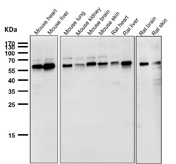
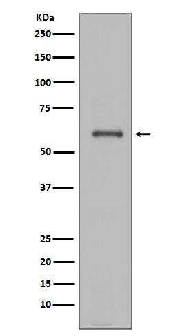
Mass (kDA):
66.519 kDA
Mouse | |
---|---|
Location: | 7|7 D3 |
Sequence: | 7; |
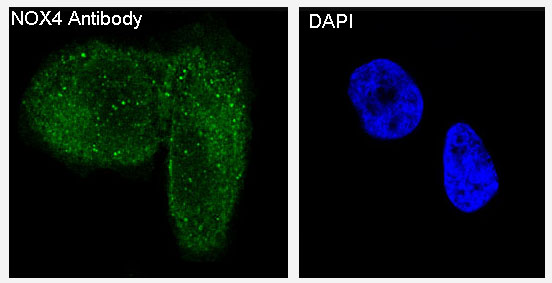
EXpressed in brain, in all layers of the cerebellum, in pyramidal cells of the Ammon horn and in Purkinje cells (at protein level). Expressed in osteoclasts, leukocytes, kidney, liver and lung.
What are the best uses of NOX4 markers? In this article, we'll look at NOX4's role as an oxygen sensor and its effects on pulmonary fibrosis, and its ability to detect apoptotic myocardial cells. It's now time for the fun part. This article will provide some information on how to get the most out of your research using NOX4 antibodies.
TGF-b stimulation causes the Nox4 gene to increase. There are many stress factors that promote NOX4 expression in cells, such as carotid balloon injury and endoplasmic and reticulum stress, hypoxia and Ischemia. There are a variety of signaling proteins and hormones that can influence the expression of Nox4 including PKCa. Furthermore, Nox4 is downregulated in response to platelet-derived growth factors and PPAR-gamma-ligands as well as serum starvation.
The intracellular location of Nox4 can influence a variety Nox4 functions, including enzyme activity, redox-sensitive cell death, and the production of ROS. The intracellular location of NOX4 has also been proven to affect the activation of various downstream signaling pathways. Nox4 is found to be present in several intracellular compartments, including the ER, PM, and cytoplasm, and is also localized to the cell membrane, periplasmic space, and nucleus.
Nox4 is one of seven members of the NADPH oxidase family. Its expression level is high in cardiac tissues, and it exhibits distinct enzymatic characteristics. Its role in the formation of reactive oxygen species is regulated through post-translational as well as transcriptional processes. The functions of NOX4 are linked to oxygen sensing and vasomotor control, cell growth, differentiation, migration and apoptonism.
NOX4's redox signaling function is vital for vascular cells. It also generates reactive oxygen species, regulates the activity of ERK1/2, and ELK1 and causes DNA damage to the nuclear genome. This NOX4 enzyme is a redox signaling one that exhibits increased activity in contrast to its isoform 1. This product is sure to be extremely useful in your lab's research.
NOX4 is an enzyme of the NOX family and acts as an oxygen sensor. It helps in the reduction of molecular oxygen to diverse ROS. It shares only 40% of the same structure as its closest relative NOX2 However, it is spliced with many variations in human cells. The protein is mRNA-positive in osteoclasts, vascular cells as well as smooth muscle cells and hemitopoietic stem-cells.
Their shared arginine residues can explain the specificity of NOX2 or NOS. The H-bond that is formed between the two NOS subunits as well as the 2'-phosphate group allow them to recognize each other. The NOX4 oxygen-sensitive marker Boster Bio's NOX4 mark recognizes both adenine and adenine and adenine is the only known molecule that can differentiate between these two groups.
NOX4 has been shown to play a role in glucose-stimulated insulin secretion suggesting that it is an essential pathway to Steroidogenesis. NOX4 also acts as an oxygen sensor in the kidney, where it regulates the synthesis of erythropoietin. NOX4 is present in human islet cells and has antimicrobial properties.
Nox4 is a key mediator of the pulmonary fibrisis. It has yet to be identified. A recent study suggests the protein could play a role in the repair and remodelling of the ASM in COPD. This study will examine the role played by NOX4 in lung function and remodeling. Further research is needed to better be aware of the way NOX4 affects the development of COPD.
The study was conducted using a small molecule known as Nox4/Nox1 inhibitor. It reduces the fibrotic response by reducing gene transcripts involved in extracellular matrix components and hypoxia-mediated vascular remodeling. The target protein is part of a complex set of transcription factors that induce the inflammatory response in fibrotic tissues, including transforming growth factor (TGF)-b1 and plasminogen activator inhibitor-1, and hypoxia-inducible factor.
Studies conducted in the past have shown that silicosis can be caused by exposure to silica in the workplace. There is no cure for silicosis, however, a natural component, Tanshinone IIA, has been found to possess anti-inflammatory, antioxidant, and anti-fibrotic properties. Tan IIA prevented the development of the disease by reducing the expression and activation of Nrf2/ARE pathways.
The NOX4 protein level rose after treatment with JHF. The drug also increased Nrf2 as well as NOX4 levels on days 7 28 and 42. This suggests that Nrf2 could play part in the therapeutic process. However, this effect was not observed in the absence of the drug rapamycin. To confirm its potential, the study will be repeated using more animal models.
Researchers have discovered that NOX4 is a biomarker used to detect myocardial cell death that is apoptotic. Boster Bio produced this marker, which can be used to detect the death of cells in the myocardium. The study utilized frozen myocardium sections. The samples were stained using an DAPI staining mixture. A laser scanning confocal microscope was used to study the slices.
In addition, to the detection of the NOX4 marker, the study also explored the role played by NOX2 in the process of apoptosis of cardiomyocytes. Researchers observed that heart tissue with higher levels of Nox4 were apoptotic and that oxidative stress promotes the death of cardiac cells. The inhibition of Nox reduced ROS production and the apoptosis of H9C2 cardiomyocytes and the results were in line with studies using other NOX inhibitors.
24 hours following SAP induction, Nox activity in myocardial tissues was determined. The expression of Nox4 protein was significantly higher in the SAP group than in the SO group, and western analysis of blots confirmed this conclusion. The results also indicated that NOX activity was implicated in injury. The NOX4 marker will allow doctors to determine apoptotic myocardial cell types.
The Nox4 protein plays an important role in vascular differentiation and fibrosis. It acts as an active regulator of TGF-beta. It also plays a role in regulating Nrf2 function in arteries. The positive effect of NOX4 on TGF-beta's expression has been demonstrated in various heart conditions from acute coronary disease to diabetes.
A NOX4 antibody is a signpost for NOX4 knockdown status. This antibody blocks cancerous breast cells from spreading. NOX4 is a part of the autophagy lysosomal pathway. There are a variety of therapeutic applications to knocking down this gene. Nox4 knockdown also decreases the production toxic metabolites in cancer cells. Here's how NOX4 knockdown works.
NADPH Oxygenase Nox4 makes superoxide within the cell. It is present in a variety of cell types including fibrocytes and vascular cell. NOX4 produces H2O2, which activates downstream signaling pathways. NOX4 also regulates the KCNK3 potassium channel, and may play a role in insulin regulation. It also is associated with atherosclerosis as well as the cardiovascular and renal complications in diabetics.
Nox4 plays a vital role in both pathological and physiological angiogenesis. In reality, knockdown of Nox4 decreases the level of the surface of VEGFR-2 which is a glycoprotein containing multiple disulfide bonds. Additionally, NOX4 knockdown inhibits the directed migration of ECs. Additionally, knockdown of NOX4 reduces the VEGFR-2 surface which alters EC angiogenic function.
PMID: 10869423 by Geiszt M., et al. Identification of renox, an NAD(P)H oxidase in kidney.
PMID: 11032835 by Shiose A., et al. A novel superoxide-producing NAD(P)H oxidase in kidney.
*More publications can be found for each product on its corresponding product page