This website uses cookies to ensure you get the best experience on our website.
- Table of Contents
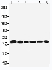
Facts about Nucleophosmin.
Connected with nucleolar ribonucleoprotein structures and bind single-stranded nucleic acids. Acts as a chaperonin for its core histones H3, H2B and H4.
Human | |
---|---|
Gene Name: | NPM1 |
Uniprot: | P06748 |
Entrez: | 4869 |
Belongs to: |
---|
nucleoplasmin family |
B23; MGC104254; NPM1; NPMnucleophosmin/nucleoplasmin family, member 1; Nucleolar phosphoprotein B23; Nucleolar protein NO38; nucleophosmin (nucleolar phosphoprotein B23, numatrin); Nucleophosmin; Numatrin
Mass (kDA):
32.575 kDA
Human | |
---|---|
Location: | 5q35.1 |
Sequence: | 5; NC_000005.10 (171387116..171410884) |
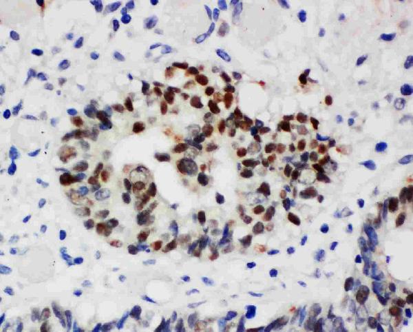
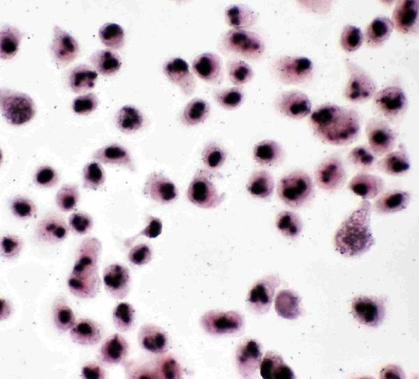
Nucleus, nucleolus. Nucleus, nucleoplasm. Cytoplasm, cytoskeleton, microtubule organizing center, centrosome. Generally nucleolar, but is translocated to the nucleoplasm in case of serum starvation or treatment with anticancer drugs. Has been found in the cytoplasm in patients with primary acute myelogenous leukemia (AML), but not with secondary AML. Can shuttle between cytoplasm and nucleus. Co- localizes with the methylated form of RPS10 in the granular component (GC) region of the nucleolus. Colocalized with nucleolin and APEX1 in nucleoli. Isoform 1 of NEK2 is required for its localization
The NPM1 marker is a member of the nucleophosmin/nucleoplasmin (NPM) family of genes. It is frequently expressed or mutated in many types of cancer. This article will explain how to utilize NPM1 in LAMs with normal Karyotype. However you choose to use NPM, you will likely discover that it's an excellent tool to use for a variety.
NPM1 is a marker gene expressed in leukemia cells. The gene expression level was determined in leukemic cells from 393 adult patients suffering from acute promyelocytic lymphoma. The NPM gene has a nucleus-restricted location in leukemic cells, and is expressed in 30 to 60 percent of leukaemia cells. The gene is expressed preferentially in monoblasts which are not yet mature.
A variety of solid tumors have been linked to the NPM1 gene family. The presence of this gene is linked to a poor prognosis. The gene is expressed in non-small cell lung cancer (NSCLC) as well as colon cancer and prostate cancer. The presence of high levels of NPM1 are associated with poor prognosis, however, it promotes cell growth. Its RNA-binding domains are remarkably conserved and can be selected, which provides further evidence of the importance of NPM1 in tumorigenesis.
The NPM1 gene is expressed in a variety of cells, including h2299 cells. The NPM1 protein is expressed by cells when the cell is grown in culture in a six-well dish. Cells were maintained for 24 hrs prior to transfection. Five mg of the expression vector that encodes wild type HA/NPM or GFP/NPM mutantA were transfected using the precipitation method using Calcium Phosphate. After that, cells were treated with 20 nM of leptomycin B.
The application of the NPM1 gene is increasing as cancer research continues to progress. The NPM1 gene is involved in ribosome biogenesis Apoptosis, as well as DNA damage repair. It is also implicated in the regulation of cell cycle, cell proliferation, and the process of apoptosis. NPM1 is also involved in the regulation of cancer cell division. NPM1 is essential in the development and maintenance of cancer-causing cell lines.
The study found that more than half of all cancers have PSs that are specific to a particular tissue. While the exact amount of PSs expressed varied across cancer types The most prevalent were those found in melanoma or thyroid cancer. Other types of cancers had a range of expression from 25 percent to 46 percent. Some PSs are found to be overexpressed in a specific type of cancer, whereas others are found in all kinds of cancer.
MDM2 overexpression could be a therapeutic target for cancer. Resistance to radiation and chemotherapy may be caused by the an overexpression of the protein. MDM2 expression in cancer may result in resistance to radiotherapy and chemotherapy. However, more studies are required to comprehend the role of this gene in the treatment of cancer. In this article, we will examine some of the main reasons why MDM2 is an ideal target for treatment of cancer.
The presence of high levels of HER2 in cancer cells make them more likely to develop metastatic or aggressive cancer. Although HER2 is not an ideal drug candidate for breast carcinoma but it has been found that it is expressed in other types of cancer. This is due to the fact that HER2 does not have a specific binding protein. However, HER2 is often overexpressed in cancer of the lungs. This protein is associated with an increased chance of developing stomach and lung cancer.
AML or acute myeloid leukemia is an hematologic malignancy heterogeneous that is characterized by the proliferation and aberrant differentiation of mature myeloid cells that are clonal. While the cause of AML remains unclear the genetic and performance-related aspects have been linked to NPM1 mutations. AML is often classified based on genetic heterogeneity. However, recent advancements in next generation sequencing have shed light onto the genetic complexity of AML.
Modern advances in the diagnosis, prognosis and treatment of AML have profoundly altered our understanding of the disease. Large-scale genomic sequencing has allowed to understand the genetic complexity of AML. It also revealed recurrent mutations within several genes. Numerous FDA-approved drugs have been developed for the treatment of mutant-specific AML. Mutations affect multiple stages of the differentiation process.
The prognosis of patients is determined by the mutation frequency of the gene. The mutational status of FLT3 has been associated with monocytic leukemia and multilineage displasia. It is associated with mutations in FAB type M4 and M5.
Of the 96 patients that had mutations in the NPM1 gene, 93% (13%) had no other alterations. Type A LAMs were affected by an NPM1 mutation, while the type B cases carried an NPM1 mutation, whereas type D patients didn't show any changes. The NPM1 mutation was absent in the other subtypes. Therefore, patients were classified as having "remaining kinds."
The frequency of the NPM1 mutation in newly diagnosed patients is high. It was also found in karyotypes with intermediate risk. We analyzed the NPM1 mutation in patients with intermediate-risk karyotypes (CBL, DNMT3A and FLT3-ITD) in this study. Additional information is available in the Online Supplementary Material.
The NPM1 marker can be utilized in LAMs to monitor and evaluate the progression of the tumor. Unlike conventional karyotypic tests, the NPM1 marker can be utilized to diagnose LAMs of any type. This marker is highly specific and sensitive, giving information on the copy number of a specific gene.
Cytogenetic analysis is utilized to determine the NPM1 status of patients who have been diagnosed with CN-AML. In older patients, the NPM1 mutation pattern is identified by an distinct genomic landscape as well as an the immunosuppressive gene signature. However the current European leukemianet genetic risk classification doesn't work well for older patients suffering from AML.
Soft tissue sarcoma refers to a form of cancer that is affecting connective tissues, such as the legs or arms. It starts as a small bump that slowly grows and spreads to surrounding tissue. Although it's not often painful, it is possible for the cancer to return if not treated promptly. The majority of people suffering from this type of cancer are at least 50 years old. Men are more likely to develop this kind of cancer than women.
Soft tissue sarcomas are classified into fifty subtypes. The subtypes are listed below and the most popular ones being listed under each body system. For instance, liposarcomas are found in fat tissue, while the gastrointestinal tumor sarcoma (GIST) starts in the digestive tract. GIST tumors are caused by mutations in the C-kit gene. This mutation isn't present in other types of. The gene is targeted by this tumor so it can shrink the tumor and reduce the risk of regrowth.
Soft tissue sarcomas could be genetically predisposed. They run in families when affected individuals have a genetic cancer-predisposition syndrome. However, if they do occur in families, it could be due to a weak immune system or other reasons. Therefore, it is crucial to seek treatment early so you can make the most effective decisions regarding your cancer. This can be accomplished with radiation therapy or surgical procedures.
DTIC was approved by the FDA in 2002 for the treatment of metastatic malignant melanoma. It has been proven to trigger subjective responses in some patients. This drug is a protodrug. It must undergo a complex metabolic process in liver before it becomes an active drug. A typical DTIC dosage is 850 to 1,000 mg/m2 every 3 weeks.
The melanomas that are in the early stages are rarely growing into lymph nodes, and therefore are a prime candidate for surgical removal. Treatment for early-stage melanomas may include surgery or other treatments. A biopsy is the initial step in the treatment of melanoma. This process involves taking a small amount of the lesion's tissue and sending it to a laboratory for analysis. Based on the results of the biopsy the treatment plan can be developed.
Immunotherapy is a different treatment option for melanomas with advanced stages. It involves the use of drugs to boost the body's immune system to recognize cancer cells. There are many types of immunotherapy, including checkpoint inhibitors. Checkpoint inhibitors aid the body's immune system to recognize cancer cells by blocking the molecule known as the checkpoint. The immune system will be more in its ability to fight melanoma cells if it targets this protein.
PMID: 2713355 by Chan W.-Y., et al. Characterization of the cDNA encoding human nucleophosmin and studies of its role in normal and abnormal growth.
PMID: 2775293 by Li X., et al. The nucleotide sequence of a human cDNA encoding the highly conserved nucleolar phosphoprotein B23.