This website uses cookies to ensure you get the best experience on our website.
- Table of Contents
1 Citations 12 Q&As


Facts about Nuclear receptor coactivator 4.

.
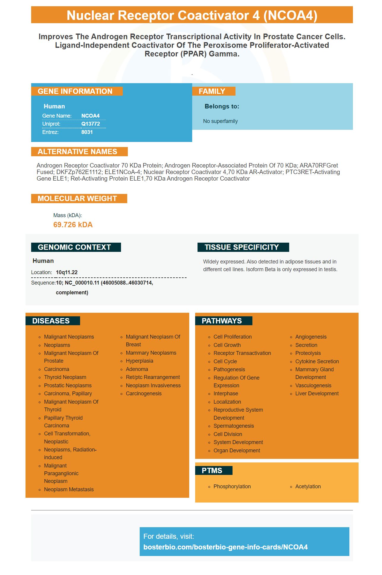
| Human | |
|---|---|
| Gene Name: | NCOA4 |
| Uniprot: | Q13772 |
| Entrez: | 8031 |

| Belongs to: |
|---|
| No superfamily |

Androgen receptor coactivator 70 kDa protein; Androgen receptor-associated protein of 70 kDa; ARA70RFGret fused; DKFZp762E1112; ELE1NCoA-4; nuclear receptor coactivator 4,70 kDa AR-activator; PTC3RET-activating gene ELE1; Ret-activating protein ELE1,70 kDa androgen receptor coactivator
Mass (kDA):
69.726 kDA

| Human | |
|---|---|
| Location: | 10q11.22 |
| Sequence: | 10; NC_000010.11 (46005088..46030714, complement) |
Widely expressed. Also detected in adipose tissues and in different cell lines. Isoform Beta is only expressed in testis.


PMID: 8643607 by Yeh S., et al. Cloning and characterization of a specific coactivator, ARA70, for the androgen receptor in human prostate cells.
PMID: 8290261 by Santoro M., et al. Molecular characterization of RET/PTC3; a novel rearranged version of the RETproto-oncogene in a human thyroid papillary carcinoma.
*More publications can be found for each product on its corresponding product page


