This website uses cookies to ensure you get the best experience on our website.
- Table of Contents
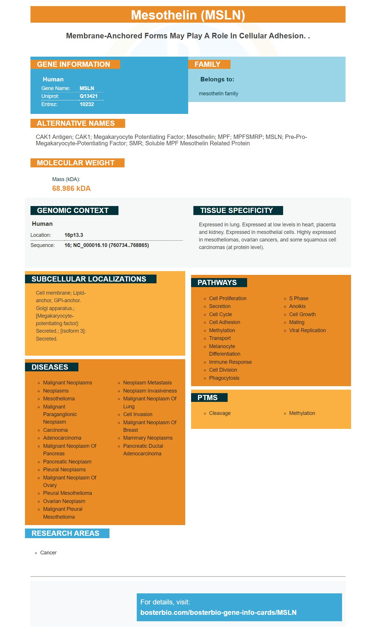
Facts about Mesothelin.
Human | |
---|---|
Gene Name: | MSLN |
Uniprot: | Q13421 |
Entrez: | 10232 |
Belongs to: |
---|
mesothelin family |
CAK1 antigen; CAK1; megakaryocyte potentiating factor; Mesothelin; MPF; MPFSMRP; MSLN; Pre-pro-megakaryocyte-potentiating factor; SMR; soluble MPF mesothelin related protein
Mass (kDA):
68.986 kDA
Human | |
---|---|
Location: | 16p13.3 |
Sequence: | 16; NC_000016.10 (760734..768865) |
Expressed in lung. Expressed at low levels in heart, placenta and kidney. Expressed in mesothelial cells. Highly expressed in mesotheliomas, ovarian cancers, and some squamous cell carcinomas (at protein level).
Cell membrane; Lipid-anchor, GPI-anchor. Golgi apparatus.; [Megakaryocyte-potentiating factor]: Secreted.; [Isoform 3]: Secreted.
When it comes to biomarkers Boster Bio has a lot to provide. In this article, we take a look at the different kinds of biomarkers and discuss their advantages and benefits as well as their uses. We also discuss picogram sensitivity ELISA kits Megakaryocyte potentiating factors, and Mesothelin. If you're seeking a biomarker for your laboratory take a look at this article to learn more about.
Immunohistochemistry (IHC) is an application of monoclonal or polyclonal antibodies to examine the distribution of a tissue antigen. It is extensively used in cancer diagnosis particularly for the detection of certain tumor antigens which are expressed in the absence of a prior signal or are increased in certain cancers. This article will review the various applications of IHC and the function of this technique in diagnostic laboratories.
IHC is traditionally performed using thin sections of tissue. For comparative analysis, small sections of tissue can be merged into one slide. The latest technology allows the automation of high-content screening to IHC. It is also possible to have samples viewed using fluorescence or light microscopy. Recent advances in technology have facilitated the collection of multiparametric IHC data and the capability to capture images.
IHC requires the highest quality dilutions. Monoclonal antibodies tend to be diluted more than polyclonal ones. Polyclonal antibodies are able to bind multiple epitopes to an antigen. Monoclonal antibodies may be more specific, but they can also give rise to background signals. Therefore monoclonal antibodies should only be employed when they are more specific.
The most important aspect in IHC/ICC tests is the choice of primary antibody. It should have high levels of specificity for the epitope of importance. Therefore, all steps of the procedure should be optimized to reduce background signals as well as to demonstrate specific staining. Initial studies must determine the best dilution for polyclonal antibodies. For polyclonal antibodies, the work concentration must be much lower than that of monoclonal antibodies.
Antibody affinity purification is an approach to improve the specificity the antibody. Polyclonal antibodies are heterogeneousand lack specificity for the antigen they immunize. In the process of immunoaffinity purification, polyclonal antibodies undergo affinity purification passing through an affinity column containing an immobilized antigen. The non-specific antibodies are eluted, while the antigen-specific polyclonal antibody is in a fixed position.
Current research is focusing on the formation and deposition of beta amyloid and paired helical filaments and the accumulation of alpha synuclein to study the neurodegenerative mechanisms. The studies on the localization of abnormal proteins have become essential to these fields, making IHC an vital tool in the diagnostic lab. IHC can be used to analyze these proteins, in addition to identifying tumor markers.
Picogram ELISA kits for MSLN include a sensitivity that measures the MSLN marker's concentration in the peritoneal fluid. The kits comprise two components: a first binding agent to the MSLN epitope and an additional antibody that is able to bind an epitope that is different. The antibodies are able to bind to each another and then identify the presence of MSLN in the sample.
These devices, methods or kits could be used in conjunction other methods for assessing the peritoneal cavities. These methods may be built on serum or dialysis fluid, or other imaging techniques like ultrasound. These tests can detect MSLN and MPF as well as other indicators of peritoneal health like inflammation and proliferative processes, and the integrity of cells.
Pourquier and VMRD both produced similar results when it came to an experiment comparing the sensitivity of several commercially available MSLN ELISA kit kits. However their sensitivity was less and their repeatability was low when compared with LSI and the BTV ELISA kits. However, the value of a homemade MSLN ELISA kit was too great to ignore.
It is not clear what function Megakaryocyte potentiating factors (TPO), plays in the growth of bone marrow. In the marrow TPO is involved in early progenitor cell development in several lineages. Animals deficient in both copies of TPO are thrombocytopenic, possess 5- to 10% Meg-CFC and have diminished erythroids and multilineage colony forming cells. TPO helps to promote the early maturation and specific amplification megakaryocyte precursors.
Both megakaryocytes that came from PMF and CTRL expressed TGFb1 receptors. TGFb1 binding was confirmed by Western analysis of blots, using anti-bactin mAb, to ensure the same loading. Both megakaryocytes derived from PMF and CTRL showed activation of pSMAD2/3, as well as Akt. The activation of PTEN was also observed in both types of cells.
Megakaryocyte potentiating factors , or MPF is an anti-mesothelin antibodies that are readily available for human consumption. It may also be called MPF, SMRP or CAK1 antigen. This protein is a major contributor to the expression by certain types of cancers and squamous-cell carcinomas, for example. These cancers could express mesothelin at high levels.
Scientists examined the role of platelets in nonhuman primates. They discovered that MGDF stimulates the growth of megakaryocytes. They also discovered that MGDF increases platelet production. Incredibly, MGDF was found to be an effective antiplatelet treatment. It is now on the market and could prove to be a successful treatment for patients suffering from immune thrombocytopenia.
The Human Mesothelin ELISA Kit has 96 wells that can be used to collect 96 samples. It is highly sensitive in detecting this protein. The kit contains antibodies against mesothelin that are specific to human MSLN. The kit is compatible with other antibodies that are used to detect other proteins in samples. The antibodies have been tested successfully in the detection of human mesothelin in clinical trials.
Human MM tumors are overexpressed for this protein. To test whether mesothelin is expressed in tumors four human MM cell lines were developed in SCID mice. The tumors that were embedded in paraffin were stained with mouse anti-mesothelin antibody. The secondary Alexa Fluor 568 antibodies were used to stain the tumors. Micrographs were captured at 400x magnification. The protein was also evaluated by Western Blot analysis using human MM cell lines as well as primary mesothelial cells from the pleural.
To assess the effects of mesothelin-based immunotherapy on the human body two studies were conducted. A multicenter phase I study compared mesothelin levels in tumor tissues with those from a single-center study. The median mesothelin level was 6.93 nM for the first study. Both studies compared mesothelin levels within tumor samples from a mesothelioma patient.
Both clinical outcomes were predicted by mesothelin levels in both these studies. Researchers concluded that mesothelin levels in tumors can predict the clinical outcome of these kinds of cancers. The dose expansion group had higher mesothelin levels than the baseline group. Mesothelioma patients had higher levels of mesothelin when compared to the control group.
PMID: 7665620 by Kojima T., et al. Molecular cloning and expression of megakaryocyte potentiating factor cDNA.
PMID: 8552591 by Chang K., et al. Molecular cloning of mesothelin, a differentiation antigen present on mesothelium, mesotheliomas, and ovarian cancers.