This website uses cookies to ensure you get the best experience on our website.
- Table of Contents
16 Q&As
5 Q&As
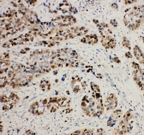
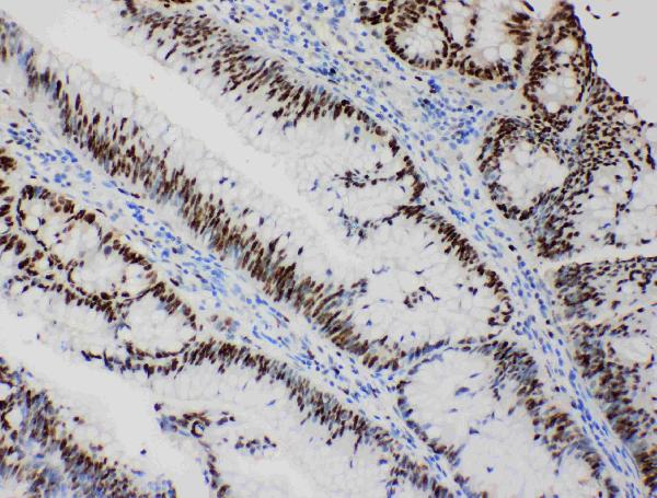
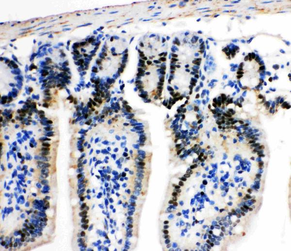
Facts about DNA replication licensing factor MCM2.
The six ATPase active sites, however, are likely to contribute differentially to the intricate helicase activity. Required for the entry in S phase and for cell division.
Human | |
---|---|
Gene Name: | MCM2 |
Uniprot: | P49736 |
Entrez: | 4171 |
Belongs to: |
---|
MCM family |
BM28; BM28MGC10606; CCNL 1; CCNL1; cdc19; CDCL1; CDCL1mitotin; D3S3194; EC 3.6.4.12; KIAA0030MITOTIN; MCM2 minichromosome maintenance deficient 2, mitotin (S. cerevisiae); MCM2; minichromosome maintenance complex component 2; minichromosome maintenance deficient (S. cerevisiae) 2 (mitotin); minichromosome maintenance deficient 2 (mitotin); Mitotin
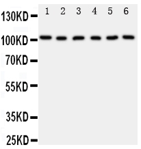
Mass (kDA):
101.896 kDA
Human | |
---|---|
Location: | 3q21.3 |
Sequence: | 3; NC_000003.12 (127598411..127622436) |
Nucleus.
This article will provide an explanation of the MCM2 marker. We will be discussing methods for cell proliferation, the Antibody in Boster Bio's MCM2 Cell Based ELISA Kit and how to apply these markers. After reading this article you'll be better able to make informed choices about your own laboratory projects.
A tumor can be detected by MCM2 staining requires a specific protein expression pattern. This marker is found in both normal and cancer tissues in the exact same way. The most beneficial results are obtained when the two markers show high levels of agreement in their expression of proteins. There are four members in the MCM2 family. If you're using the MCM2 marker to detect a tumor marker, you need to determine which one is appropriate for your situation.
MCM2 is expressed at the time of cell-cycle initiation and continues to be expressed throughout the cycle of the cell. MCM2 also shows immunoreactivity in all active phases of the cell-cycle, excluding G0. The expression of Mcm2 is a useful biomarker for the potential for reproduction. It is used to detect the presence of precancer-related cells and is associated with precancer-related growth.
Researchers have successfully isolated MCM2 from yeast to mimic the pre-RC process. When loaded with DNA, MCM2 forms a double hexamer. This is crucial for understanding DNA licensing and replication. While the MCM2 marker isn't widely available, it does offer numerous advantages. Its high level of specificity makes it an extremely desirable alternative to Ki-67.
The MCM2 protein is a proliferative-associated minichromosome maintenance protein that regulates cell proliferation and recruits replication machinery. It is often expressed up-regulated in cells that are proliferating. It is believed that Mcm proteins are more specific than Ki-67/MIB-1 for determining tumor growth. Although there are positive results for Mcm2 and gliomas , more studies are needed to confirm these findings. The Mcm2 proliferative index has a strong correlation mitotic and survival indexes, and it has a strong relationship with the risk of recurrence.
Tissue microarrays were developed using samples from 249 breast cancer patients. The samples were then used to verify the MCM2 expression thresholds. The positive correlation between the MCM2 marker and the other four markers was found by using three scoring methods. It is crucial to check breast tissue for the MCM2 marker even if it's not found in breast cancer.
Several methods are available to measure cell proliferation with the MCM2 marker. These methods combine biochemistry and instrumentation with sensitive reagents which allow precise measurements of specific cells in complex models. Multiparametric cell proliferation analysis can also be performed using changes in morphology and indicators of additional cells' events. Multiparametric analysis can be a powerful tool for basic research , and could be used to develop and discover new drugs.
The MCM2 marker can also help differentiate between normal cells exhibiting abnormal growth. It is present during the early G1 phase and is relevant to determine the behavior of tumors. Ki-67 tests can also be used to predict the growth of various tumors. These methods may yield contradictory results. However, they are beneficial in identifying tumors and are typically used in tandem.
The AurkbER Cre/+ mouse is a promising instrument for tracking the lineage of cells in mice. It can be crossed with confetti mouse to track the development of clonogenic neural cells, trace rare stem cells derivatives, as well as detect the progression of diseases. This allows for diagnostics based on cells like tumor regenerative therapies.
MCM2 is one six members of the MCM family, which plays a significant role in DNA replication. Furthermore, it has distinct cell-specific localization patterns, making it a better marker of proliferation than Ki-67/MIB-1. However studies on Mcm2 in gliomas showed inconsistent results, however, the proliferative index of Mcm2 in these tumors demonstrated a significant correlation with the mitotic index and survival and recurrence.
The MCM2 antibody is extremely specific against cell-specific secondary antibodies, and compatible with other cell-specific primary antibodies. The effectiveness of the antibody was confirmed using Western Blot and qRT-PCR of LUSC cells. Additionally, MCM2 downregulation diminished the viability of SK MES-1 cells, decreased the number of colonies and increased the proportion of cells in the G0/G1 phase.
In addition to these methods, MCM2 expression in lung cancer tissue can be assessed by immunohistochemical analysis. The MCM2 mRNA levels were increased in the two human LUSC cell varieties. This was further confirmed by the high levels of MCM2 expression in lung cancers. These findings are significant for the future of cancer research. These techniques could be used to detect early stages of cancer.
MCM2 immunostaining is linked to lower survival rates in astrocytomas with grade II. However, there is no evidence to suggest that Mcm2 immunostaining is superior to Ki67/MIB-1 in human astrocytomas. There are some trends that suggest lower survival rates for patients with high levels of Mcm2. To confirm Mcm2's role in cancer, more research is required.
Boster Bio's ELISA Kit MCM2 uses an IHC optimized polyclonal antibody. This allows researchers to detect multiple biological markers at picogram-level sensitivity. Boster Bio's antibodies include more that 12,000 antibodies that have been tested for use with cell-based assays WB, Flow, and. To ensure high affinity, these antibodies have been thoroughly tested against 250 tissues. Boster Bio's ELISA kit is equipped with advanced immunogen technologies such as Picoband or Supervision. These tools let researchers quickly and effortlessly conduct quantitative testing using their antibodies against a range of cell lines or tissues.
The MCM2 Cell Based ELISA Kit can detect MCM2. Researchers can analyze the effects of various stimulation conditions on MCM2 expression in various cell lines. Anti-MCM2 antibodies are able to capture the protein and are recognized by HRP-conjugated primary antibodies. Secondary antibodies are conjugated with HRP enzymes in order to initiate the colorimetric reaction. The MCM2 Cell-Based ELISA Kit also offers a variety of normalization methods.
The results of this research support the theoretical model that the antibody attaches to the cell's surface. This model is the basis for more effective therapies and timely quantitative imaging probes. While the findings of the study are not conclusive but these findings are in favor of the theoretical model. This model could be used as a base for accurate ELISA testing, if further refined. It can also be used to track antigen expression in different cancer cells.
Two monoclonal monoclonal antibodies are employed in sandwich assays. They have different epitopes. The first antibody is responsible for removing the protein from the sample, while the second antibody provides the signal that shows the presence of the target. Sandwich assay is highly sensitive and has many benefits. A standard curve can be used to analyze the signal from a single sample.
The VHH26 Clone was fully active in ethanol, 0.5% 2-ME, and 0.5 M GuHCl. It was capable of detecting gliadins from samples that were previously rejected by conventional ELISA methods. This antibody is a good platform for quantitative protein determination. It is compatible with a variety of other antigens.
The Antibody used in the Boster Bio's MCM2 Cell Based ELISA Kit is made in the laboratory from an anti-parvalbumin-like polyclonal antibody. It is extremely specific for the southern hemisphere fish and shows strong phylogenetic ties with the antibody. However this anti-parvalbumin antibodies might not be sufficient to identify fish species.
PMID: 8175912 by Todorov I.T., et al. A human nuclear protein with sequence homology to a family of early S phase proteins is required for entry into S phase and for cell division.
PMID: 8258304 by Mincheva A., et al. The human gene for nuclear protein BM28 (CDCL1), a new member of the early S-phase family of proteins, maps to chromosome band 3q21.