This website uses cookies to ensure you get the best experience on our website.
- Table of Contents
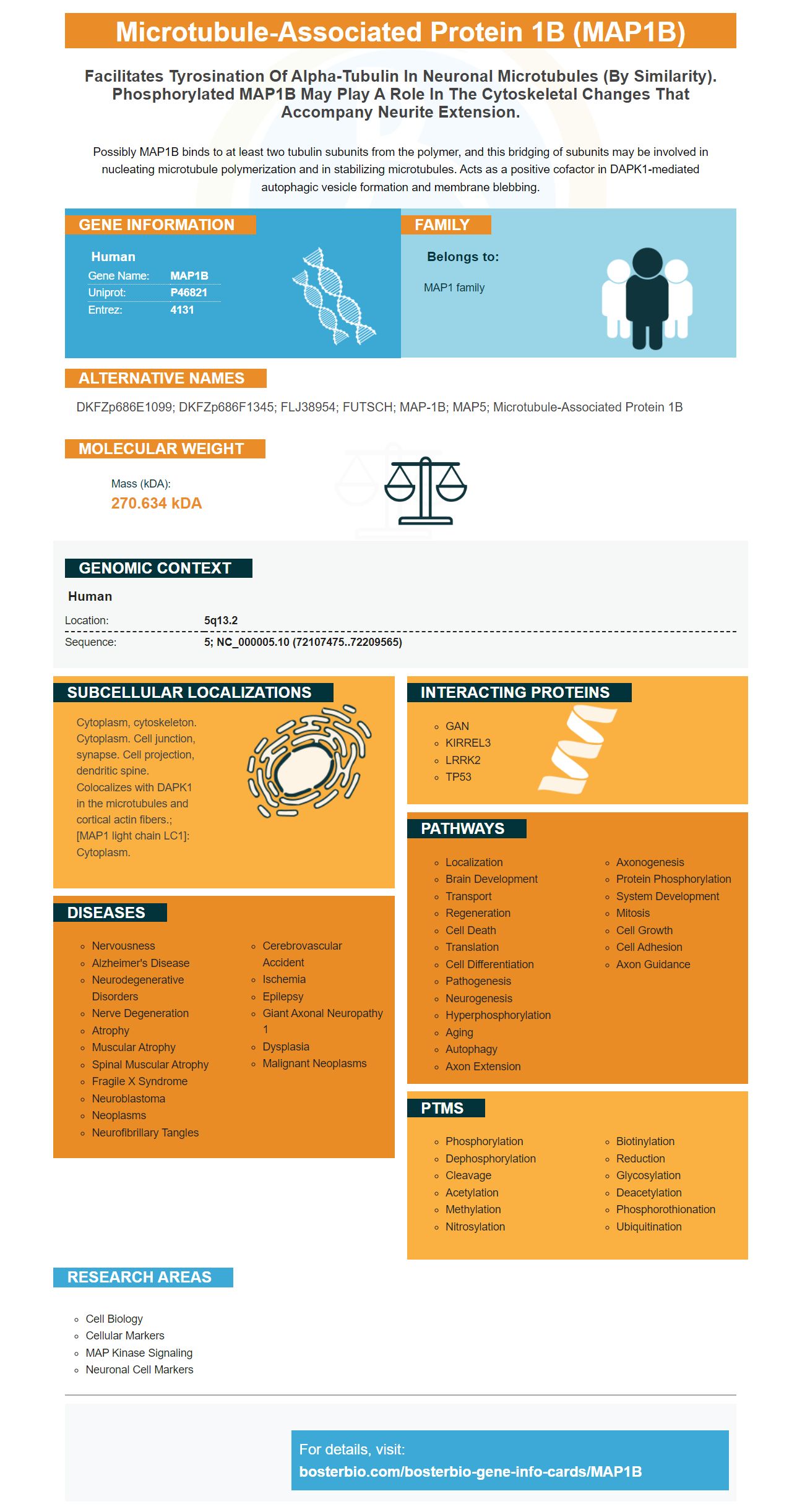
Facts about Microtubule-associated protein 1B.
Possibly MAP1B binds to at least two tubulin subunits from the polymer, and this bridging of subunits may be involved in nucleating microtubule polymerization and in stabilizing microtubules. Acts as a positive cofactor in DAPK1-mediated autophagic vesicle formation and membrane blebbing.
Human | |
---|---|
Gene Name: | MAP1B |
Uniprot: | P46821 |
Entrez: | 4131 |
Belongs to: |
---|
MAP1 family |
DKFZp686E1099; DKFZp686F1345; FLJ38954; FUTSCH; MAP-1B; MAP5; microtubule-associated protein 1B
Mass (kDA):
270.634 kDA
Human | |
---|---|
Location: | 5q13.2 |
Sequence: | 5; NC_000005.10 (72107475..72209565) |
Cytoplasm, cytoskeleton. Cytoplasm. Cell junction, synapse. Cell projection, dendritic spine. Colocalizes with DAPK1 in the microtubules and cortical actin fibers.; [MAP1 light chain LC1]: Cytoplasm.
The MAP1B marker can be used for research purposes in many different areas. This article will discuss how MAP1B is expressed in the glomerulogenesis process, immunolabeling, synthesis, as well as applications. These are all topics Boster scientists should be aware of. These methods can be applied worldwide. These are the best uses for the MAP1B marker in research.
MAP1B has been proposed as an essential protein during glomerular development. Its knockout mice show a phenotype that cannot be explained. Here, we report on the analysis of MAP1B expression during glomerulogenesis in mice. We show that podocytes express MAP1B. It is interesting to note that podocytes express CFP when they are stimulated by nephrin.
We found a heterozygous mutation within the MAP1B gene (c.4198A–G). This mutation is responsible for deficient phosphorylation and instability. Map1B, which is also found in the kidney, is also associated microtubules. The p.1400S-G mutation has a significant negative effect on microtubule assembly, and we are investigating the role of MAP1B in this process.
We also showed that glomerular permeability was affected by MAP1B deficiency. It is able to induce proteinuric acid in the kidney of the rat by inhibiting expression P0. A study by Pavenstadt et al. Pavenstadt et al. showed that MAP1B p.1400S–G is associated with altered dynamics of microtubules, which could hinder axon elongation.
MAP1B can be expressed during glomerulogenesis. Progestogenic treatment did not alter the expression of MAP1B during this process. Progestogenic hormones also did not affect the uptake and use of thymidine labeled RNA. Furthermore, PR expression was not correlated with DNA polymerase activity, suggesting that it is a splicing factor of MAP1B.
Immunolabeling MAP1B revealed that mRNA was enriched in dense MT cytoskeletons in glomeruli as well as the cell body. The arrowheads on morphological images highlight the primary process and foot processes. Meanwhile, the nucleus was highlighted by 200 nm. In contrast, MAP1B expression could only be detected in a small section of human kidney.
The MT cytoskeleton, which is found in glomerulocytes, is highly arborized. Their major processes have many similarities with the dendrites of neuronal cells. The MT cytoskeleton is regulated by the phosphorylation of microtubule-associated proteins (MAP1B). Depletion of MAP1B inhibits the formation these processes. Therefore, MAP1B depletion may be a factor in the regulation of the function of spiral ganglion neurons.
MAP1B is a protein which is involved in maintaining the microtubule dynamics. MAP1B mutations result in reduced levels of the protein and decreased phosphorylation. MAP1B mutations resulted in cells with altered electrophysiological properties as well as disturbed microtubule dynamics. These cells were saved by genetic correction using CRISPR/Cas9 technology, which restored their hearing abilities. These experiments also revealed that MAP1B is involved with hearing and auditory function.
This study also showed that immunolabeling MAP1B confirms that this protein is present in podocytes' dense MT cytoskeleton. Immunolabeling MAP1B confirms that this protein is found in podocytes during glomerular differentiation. This occurs as the cortex becomes the medulla. MAP1B is also found to co-localize with MT bundles in the rat kidney.
In neuronal somata, cortical layer VI and aged rats, MAP1B was found to have an aberrant immunolabeling. This observation indicates a progressive loss or gene activity with old age. Neuronal Plasticity has been implicated by MAP1B. While these findings are still in the early stages of investigation, they have significant implications for research and therapy. The findings suggest that immunolabeling of MAP1B is an important step in defining glomerular function.
To carry out immunolabeling experiments, we used primary antibodies from Beat M. Riederer, University of Lausanne. Secondary antibodies were used for this research: goat anti-rabbit IgG; MAP1B BR18 antibody; and mouse anti–mouse IgG. The sections were embedded in Durcopan, and then analysed on a Zeiss Leo 906TEM.
MAP1B was also highly expressed in spiral ganglion neurons, a type of neuron that contains the gene. The gene was compared with b-actin levels in different tissues. The MAP1B expression levels were then normalized to b–actin in three separate experiments. The resulting images were then examined by immunofluorescence. Because MAP1B is abundant in these neurons, it is believed to be involved in the function of spiral ganglion neurons.
Mutations in MAP1B are the cause of otosensory neuron dysfunction. MAP1B is expressed at high levels in the neuron system and at low levels in other tissues. Using this mutant mouse model, iPSC-derived cell lines were transformed into OSN-like cells. The morphology, phosphorylation activity and electrophysiology of these iPSCs was determined.
Synthesis of MAP1B marker by boster bio. This gene encodes the mCherry MAP1B marker, which is a highly specific marker for podocytes. The protein is linked to the podocyte MT skeleton. The mCherry tagging on the MAP1B marker permits detection of the protein by antibody-mediated immunofluorescence.
The MT cytoskeleton has a complex structure that has been studied well on a morphological scale, but has not been thoroughly investigated in the context glomerular filtration. The MT cytoskeleton, which shares many similarities with the MT-bundles in neurons, is an important part for cell process outgrowth. The assembly of MT bundles during glomerular filtration is affected by MAP1B depletion.
The MAP1B protein has a diverse role in the body's cellular function. The polyclonal and monoclonal antibodies that are used for biochemical studies react with MAP1LC3C in various animal samples. Boster Bio uses rabbit and mouse as their model organisms. This protein could also play a role in the cellular process known to be aggrephagy.
Mutations that cause axodendritic disorders in PD may have an impact on the body's immune system. MAP1B expression has been linked to a reduction in axonal regeneration in PD. The mutation that causes this problem for mice may cause additional signaling, independent of ringer. This could result in reduced expression of tau.
Among many studies done on the MAP1B indicator, results show a positive relationship between IQ & FSIQ. A paired test showed that MAP1B carrier had a higher intelligence than non-carriers. This was due to lower scores on the Block Design subtest. Moreover, a greater VIQ was seen in disorders with NVLD-like symptoms, such as autism spectrum disorder and Fragile X syndrome. On the other hand, a lower PIQ was observed in disorders such as agenesis of corpus callosum and Fragile X syndrome.
MAP1B has a close association with the podocyte-MT skeleton. The podocyte's MT skeleton is closely associated with the protein's gene expression. This gene could play a crucial role in many human diseases. It is likely that MAP1B will be used in biomedical research as well as clinical medicine. These applications of MAP1B are expected to increase.
To find out the effects of MAP1B on kidney, we used mouse kidneys. The kidneys had to be dissected and cut into sections measuring 50 mm in thickness. MAP1B BR18 antibody (1:150) was applied to the tissues and stained with Nanoprobes' DAPI-conjugated goat anti-rabbit secondary antibody (NAB8926). These sections were embedded using Durcopan. The Zeiss Leo 906TEM analysed them.
MAP1B LoF variants have smaller CC and brain-wide WM volume. These findings provided insights into the molecular mechanisms underlying human neurodegenerative disorders. This gene is essential in regulating MAP1B in the brain. It also influences expression of other proteins and genes. It is useful in many neurodegenerative disorders. MAP1B is an important intermediate in the formation of the central nervous system.
The MAP1B protein is present in podocytes during differentiation. MAP1B HC co-localizes with the MT bundles of podocytes. Positive controls for this research included the lysates hek-293T cells and brain lysates. These cells can be used for immunofluorescence microscopy. MT fusion occurs when the MAP1B HC & MT bundleds co-localize.
PMID: 7806212 by Lien L.L., et al. Cloning of human microtubule-associated protein 1B and the identification of a related gene on chromosome 15.
PMID: 12684070 by Dergunova L.V., et al. Hmob3 brain-specific sequence is a part of phylogenetically conserved human MAP1B gene 3'-untranslated region.