This website uses cookies to ensure you get the best experience on our website.
- Table of Contents
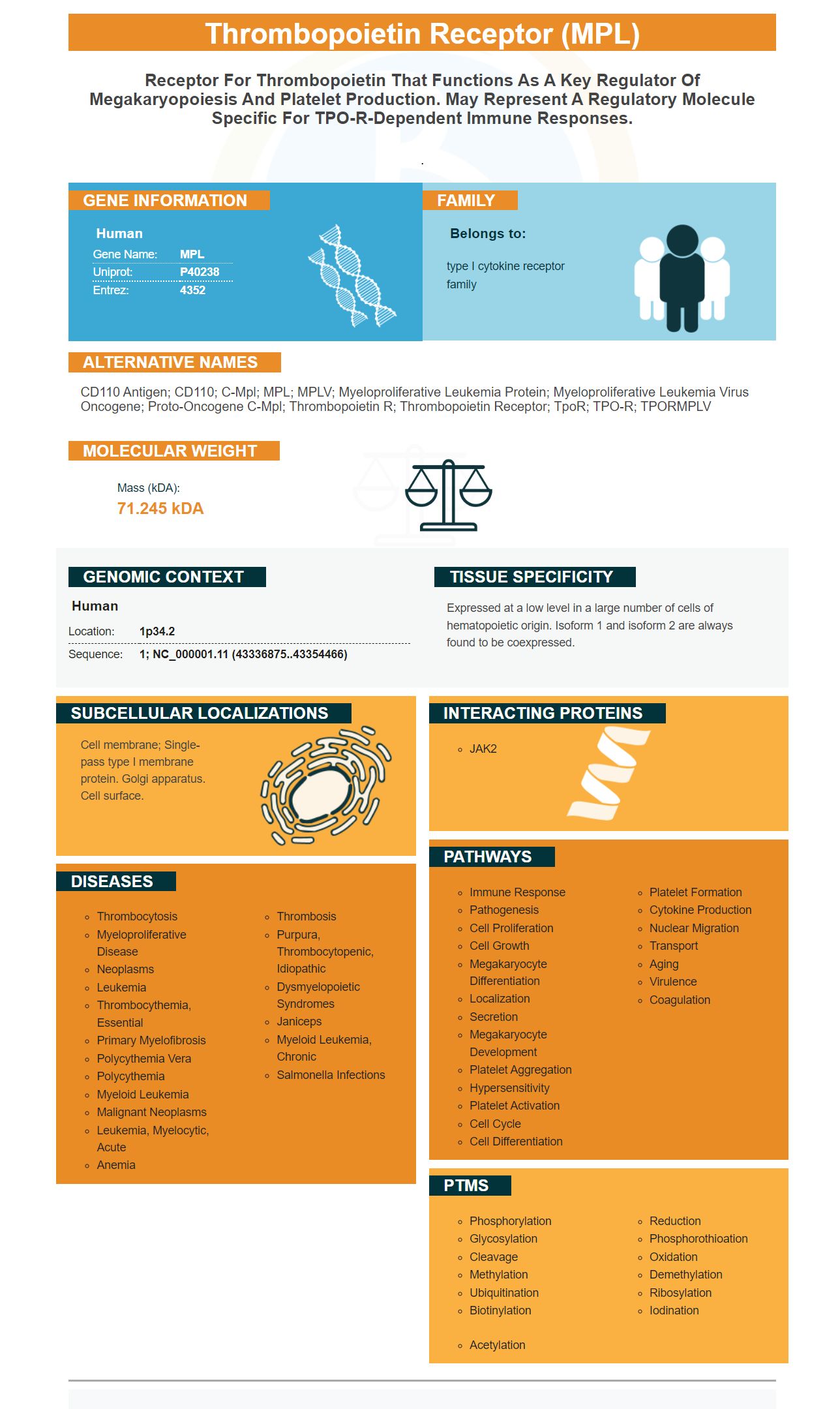
Facts about Thrombopoietin receptor.
.
Human | |
---|---|
Gene Name: | MPL |
Uniprot: | P40238 |
Entrez: | 4352 |
Belongs to: |
---|
type I cytokine receptor family |
CD110 antigen; CD110; c-Mpl; MPL; MPLV; Myeloproliferative leukemia protein; myeloproliferative leukemia virus oncogene; Proto-oncogene c-Mpl; Thrombopoietin R; thrombopoietin receptor; TpoR; TPO-R; TPORMPLV
Mass (kDA):
71.245 kDA
Human | |
---|---|
Location: | 1p34.2 |
Sequence: | 1; NC_000001.11 (43336875..43354466) |
Expressed at a low level in a large number of cells of hematopoietic origin. Isoform 1 and isoform 2 are always found to be coexpressed.
Cell membrane; Single-pass type I membrane protein. Golgi apparatus. Cell surface.
The MPL Marker is a tool that scientists use to track their progress. What exactly is the MPL Marker? And what are its best uses. Let's find out. First, this is an anti Cytokeratin Pan (Epithelial marker) antibody. We will then learn how we can handle this marker. This antibody is very useful for scientists around the world.
Cytokeratin, Pan (Epithelium-Associated Membrane) is a cell-surface protein present in several tissues, including epithelia, hematopoietic stem cells, and neuronal cells. It is also present within the human body. It has been used to distinguish between different neoplasms. It is sensitive to Cytokeratins and used to distinguish between normal and cancerous cells.
Cytokeratin, Pan (Epithelium-Associated Membrane Protein) is a subfamily of intermediate filament proteins, and it is highly varied in its biochemical composition. At least 20 different polypeptides can be found in human epithelia. Their molecular weights range from 40 to 68kDa to 64 kDa. Cytokeratin, Pan (Epithelium-Associated Membrane Protein) is present in both benign and malignant tissues.
Cytokeratin, Pan (Epithelium-Associated Membrane Protein) Antibody is useful in diagnosing various types of cancers. It can identify single or grouped carcinoma cells as well micrometastases in paraaortic and para-aortic lymph nodes. It has a cytoplasmic immunoreaction that is highly specific for cancer cells. It can detect carcinoma cells even in tissue that has occult or absent tumor cells.
Cytokeratin, Pan (Epithelium-Associated Membrane) is expressed in pairs and has been used in a variety of applications. In many applications, CK1 & CK10 pair up with each other. Pan (Epithelial Marker). Antibody can be used to detect CK1 and CK2 in tissues.
Antibodies are used in immunohistochemistry in a variety of ways, including the use of antiCK, Pan, or EpCAM. These antibodies have proven to be invaluable in clinical diagnostics. They are especially useful in studies involving human tissue. You can also use the antibodies to culture alidifferentiated cells. BosterBio's Polyclonal Antibody is a good choice if you are looking for an anti-CK, Pan (Epithelial Marker), Antibody.
This anti-CK7 antigen can be used in conjunction H&E morphology to help determine if there are epithelial markers present in bowel cancer. Flat bowel mucosa is a condition that anti-CK7 antibodies can help diagnose. It can also be used to detect epithelial dysplasia.
A patient with suspected fibroblastic Os presented with swelling of the left maxilla. The swelling was larger and more measured. The patient's pathology revealed a population of cytokeratin AE1/Three-positive spindled cells in a storiform sample. Fluorescent, in situ hybridization confirmed that this was a fibroblastic osarcoma. Furthermore, the patient's medical history was complete.
Matplotlib C++ is a lightweight wrapper of the Python matplotlib Library. The code is available at GitHub. This C++ library makes it easy to work on MPL data. It also supports basic MPL commands. The code can be accessed in The Docs, as are documentation and examples. There are also useful functions to deal with MPL markers.
Matplotlib's scatterplot function allows you to plot scatterplots by using the scatterplot function. It can also be used to plot scatterplots, which are essentially graphs composed of square regions. Each square area is bound by a number. Each marker indicates a different area. Divide the length by 2, to calculate the area. To learn more, use Stack Overflow. The plot becomes difficult to read when the markers are too small.
Patients with multiple diseases are becoming more interested in the MPV-marker. Researchers have found that the expression of this protein is associated to a neoplastic mutation that alters platelet parameters. Recent research has evaluated platelet counts and the MPV/PLT relationship in patients with advanced hepatocellular malignancy. Patients with carcinoma had higher levels of MPV than healthy people, and were more likely to have synchronous liver metastases.
The MPL marker has many uses, including scatter and plot. Plot, scatter, and errorbar use it. The marker center is located at (0.0). The marker size is normalized so it fits within the unit cell. Markers are a regular polygon that has numsides sides, or a symbol like a star or an asterisk. The data is not available if 'None’ is set.
PMID: 1608974 by Vigon I., et al. Molecular cloning and characterization of MPL, the human homolog of the v-mpl oncogene: identification of a member of the hematopoietic growth factor receptor superfamily.
PMID: 8020956 by Mignotte V., et al. Structure and transcription of the human c-mpl gene (MPL).