This website uses cookies to ensure you get the best experience on our website.
- Table of Contents
2 Citations 16 Q&As
Facts about Induced myeloid leukemia cell differentiation protein Mcl-1.
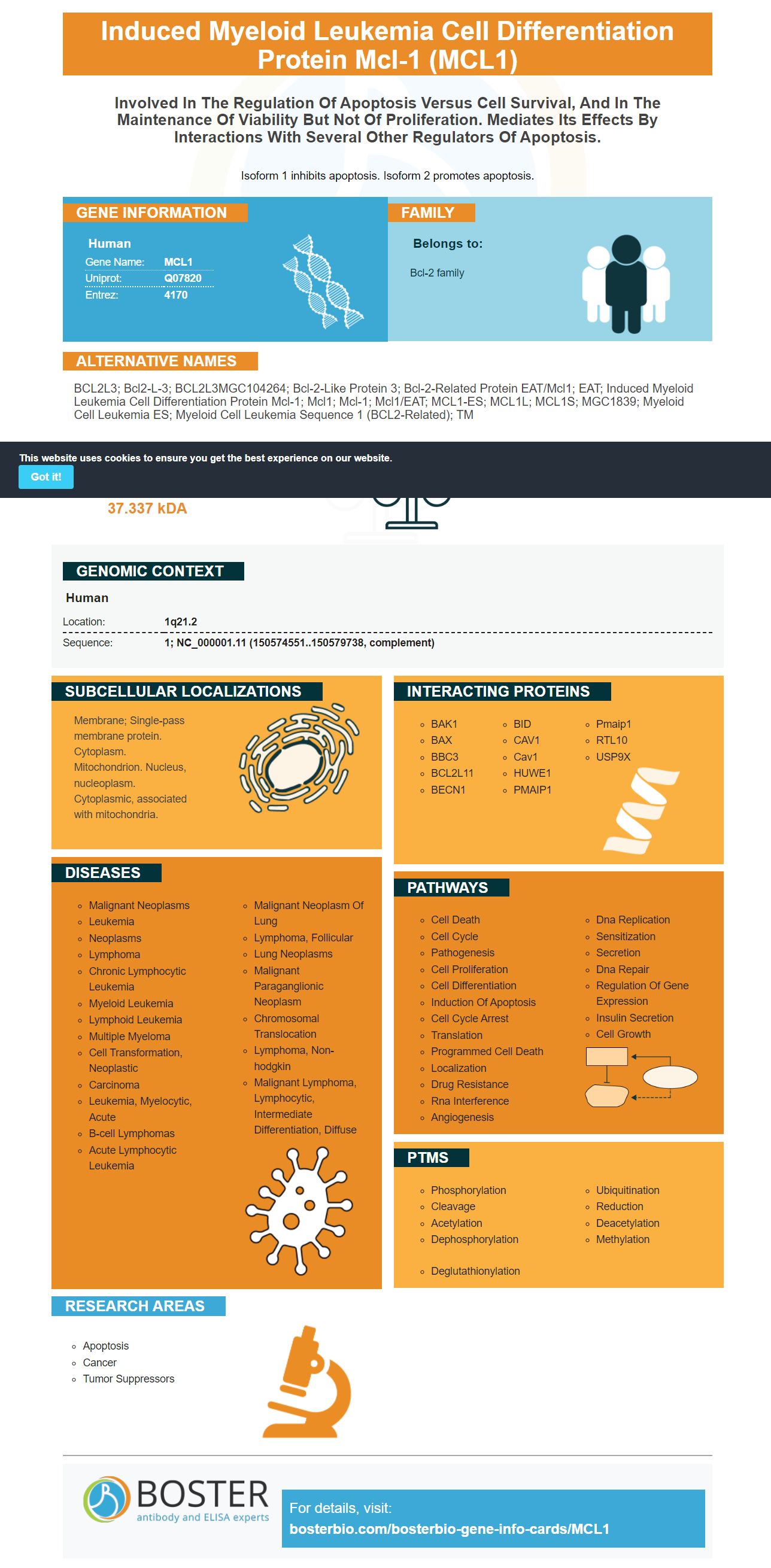
Isoform 1 inhibits apoptosis. Isoform 2 promotes apoptosis.
Human | |
---|---|
Gene Name: | MCL1 |
Uniprot: | Q07820 |
Entrez: | 4170 |
Belongs to: |
---|
Bcl-2 family |
BCL2L3; bcl2-L-3; BCL2L3MGC104264; Bcl-2-like protein 3; Bcl-2-related protein EAT/mcl1; EAT; induced myeloid leukemia cell differentiation protein Mcl-1; Mcl1; Mcl-1; mcl1/EAT; MCL1-ES; MCL1L; MCL1S; MGC1839; myeloid cell leukemia ES; myeloid cell leukemia sequence 1 (BCL2-related); TM
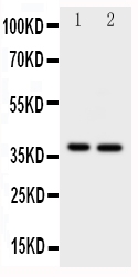
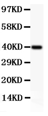
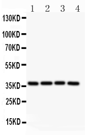
Mass (kDA):
37.337 kDA
Human | |
---|---|
Location: | 1q21.2 |
Sequence: | 1; NC_000001.11 (150574551..150579738, complement) |
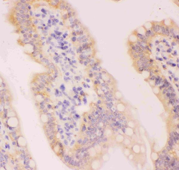
Membrane; Single-pass membrane protein. Cytoplasm. Mitochondrion. Nucleus, nucleoplasm. Cytoplasmic, associated with mitochondria.
This article will examine the role of MCL1 in cell growth and health. We will also discuss its potential as a biomarker for anti-tumor therapy, and its use as a target for proteolysis-targeting chimeras. This article is for those who are in search of a new biomarker. This article has everything you need to know about starting.
MCL1 is a member in the Bcl-2 family , and controls various cell processes. It plays an important role in cell division and repair of double-strand DNA breaks mitochondrial dynamics, and bioenergetic metabolism. Moreover, overexpression of this protein is associated with resistance to targeted and conventional cancer treatments. We will now discuss the vital roles of MCL1 within the cell cycle, and then discuss its role in the fight against cancer.
There has been a lot of debate about the role played by Mcl-1 for the process of apoptosis. Mcl-1's pro-survival role is linked with the decrease in its expression by various forms of stress. Cytokine deprivation, UV radiations and prolonged mitotic arrest are all common examples of these stressors. Mcl1 is an atomic "timer" for cell death. Its decreased expression triggers cell death.
Mcl-1 is also highly regulated at multiple levels, and is related to the death of cells. Its expression is tightly controlled at various levels, involving transcription, post-transcriptional process as well as proteasomal degradation. The protein is then targeted for degradation by proteasomes, which leads to cell death. This mechanism has been extensively researched over the past decade. In addition to this, Mcl-1 is also involved in various metabolic pathways.
Additionally, the an overexpression of MCL1 inhibits cell cycle progression through the S phase. In vitro, MCL1-/blastocysts from mice were not able to hatch in vitro. MCL1 is a gene that blocks cell cycle progression through S phases, however it still has its antiapoptotic as well as DNA-synthesis functions. Additionally MCL1 negatively regulates the protein p18 that helps to promote cell cycle entry and G1/S transition. Additionally, MCL1 interacts with PCNA, the DNA sliding clamp that acts as a cofactor for DNA polymerase d in S phase.
MCL1 regulates mitochondria's ultrastructure which allows for fusion and fission. It also interacts with two GTPases OPA-1, and DRP1. DRP1 creates an oligomeric ring around mitochondria at the OMM which facilitates fission, which is beneficial to the survival of cells. OPA-1 is the mediator of mitochondrial fusion at the MIM.
MCL1's anti-apoptotic activity regulates various cells' processes, including cell differentiation and progress. MCL1 also plays a role in the repair of double-strand DNA and mitochondrial dynamics, as well as bioenergetic metabolic metabolism. However, the roles of these proteins aren't fully understood. We will be focusing on the functions of the cell MCL1 & BCL2 in the current study.
Recent studies have shown that MCL1 also interacts with rBH3, which might play an important role in regulating cell fate. More research is required to determine if these proteins possess similar roles. These interactions could also be relevant to MCL1 matrixforms. This pathway is extremely complex and we are only beginning to grasp its intricate details. Our goal is to better know the molecular mechanism behind it and the molecular mechanisms that regulate them.
MCL1's antiapoptotic activity is essential for the survival and differentiation of a variety of cells including neurons. MCL1 is involved in regulation of cell death and cell formation during hematopoiesis. Moreover, this enzyme is crucial for the differentiation of blood cells from stem cells. The deletion of MCL1 causes impairment to this process and causes the loss of hematopoietic stem cells.
MCL1 has been involved in autophagy. It is a method for recycling and degrading intracellular material. The function of MCL1's cells is contingent on the context within which it is located. MCL1 targets mitochondrial pathways to block autophagosome production. One possible mechanism behind the induction of autophagy by preeclamptic uterine placentas is through the interaction between MCL1 and BECLIN1.
One of the major issues when evaluating treatments for cancer is that there is no single marker that can reliably determine the effectiveness of any specific treatment. Researchers are currently investigating the function of sCD163 as an anti-tumor treatment. It is present in tumor cells of patients with advanced melanoma , who were previously untreated. It is also known that it can alter the response to ICIs,integrin-dependent Kinases.
Many cells in the body produce IL-6, which includes cancer cells. It is crucial to tumor progression, inhibits the death of cancerous cells, and also promotes angiogenesis. In one study, patients suffering from advanced melanoma who had high levels of IL-6 levels in their blood had a shorter OS when compared to those treated with IL-2-based immunotherapy. So far, the serum concentration of IL-6 has not been associated with response to immunotherapy. However CRP, which is able to be controlled by IL-6 has been demonstrated to predict outcomes in patients who have received ICIs.
ICI treatment prediction is possible using several predictive biomarkers. However, multiple biomarkers need to be considered when deciding on which ICI to use. Since ICIs can trigger complex immune responses, the discovery of new biomarkers can provide mechanistic insights into the effects of these drugs on the immune system. This will lead to better anti-tumor therapies. It is vital that we do not forget that the process of creating biomarkers that are new is still in its early stages.
The NCC-GP150 study contains technical as well as virtual clinical validation of the GP150 panel. The NCC-GP150 panel has been validated using WES data collected from the TCGA. The panel has been evaluated against other gene panels like MSK IMPACT and PlasmaSELECT64 as well as a public NSCLC cohort. Multiple studies have validated the NCC-GP150-based TMB.
Proteolysis-targeting chimeras (PROTACs) are small molecules that recruit E3 ligase and the protein of interest to undergo polyubiquitination and subsequent degradation by the proteasome system. PROTACs remain highly empirical due to the complexity of the degradation process. A more rational approach to the development of PROTACs would involve profiling step-by step in the ubiquitin-proteasome degradation process by using biophysical assays.
One such PROTAC molecule is a POI-ligand which is linked to the E3 Ligase via the linker composed of five to 15 carbon atoms. Its mechanism involves proximity-induced ubiquitination in POI, which is later degraded by the 26S proteasome. This mechanism was discussed in a recent x-ray crystal structure of the POI PROTAC-E3 ternary structure.
Similar to this, PROTACs targeting the BRD4 kinase, TKD1, were designed to block BRD4 degradation. PROTACs with this inhibitor blocked BRD4 expression in MV4;11 leukemia cells in the laboratory. In addition, PROTACs targeting BRD4 were able to inhibit the growth of p53-wild type cancer cells in both human and mouse samples.
A better understanding of how tumor cells escape death is driving the development of new anti-tumor drugs. Cell death escape is closely connected to the dysregulation of proteins from the Bcl-2 family. New possibilities have been opened up in the treatment and prevention of cancer thanks to the discovery of novel anti-tumor drugs that are based on MCL1 inhibition. To maximize the efficacy of this novel drug class it is necessary to understand the full extent of the role of MCL-1.
Because of the numerous implications of MCL-1's role in the advancement of cancer survival, MCL-1 inhibitors can improve the outcome of patients suffering from metastatic or advanced Melanoma. It is possible to combine MCL-1 inhibitors and MEK inhibitors, especially for KRAS mutations in NSCLC. Additionally, a thorough understanding of the BCL-2 family's dependencies will help in the development of true precision medicine.
In addition to the anti-tumor properties of MCL-1 inhibitors and thrombocytopenia, by pharmacologically activating MCL1, it can cause thrombocytopenia. Proteolysis-targeting chimeras are another promising approach to target MCL1.
Inhibiting MCL-1 which is a molecule that blocks the apoptotic machine, could be a significant treatment option for patients suffering from blood cancers that are hematological. There are several potent and specific MCL-1 inhibitors currently being developed. They are not intended to replace other targeted therapies, but can be beneficial additions to biomarker-driven therapies. They should be used in conjunction with other targeted treatments.
There are a variety of approaches that have been developed to boost the degradation of MCL-1 by targeting USP9X. These therapies focus on the MCL-1 pathway to eradicate cancer cells. Some of these agents can also reverse MCL-1 resistance. One of the drugs, cobimetinib is able to reverse MCL-1 inhibition. Numerous studies have demonstrated that MCL-1 is the key for a new type of anti-tumor agent.
PMID: 7682708 by Kozopas K.M., et al. MCL1, a gene expressed in programmed myeloid cell differentiation, has sequence similarity to BCL2.
PMID: 8790944 by Umezawa A., et al. Induction of mcl1/EAT, Bcl-2 related gene, by retinoic acid or heat shock in the human embryonal carcinoma cells, NCR-G3.
*More publications can be found for each product on its corresponding product page