This website uses cookies to ensure you get the best experience on our website.
- Table of Contents
9 Citations 9 Q&As
2 Citations 6 Q&As
4 Citations
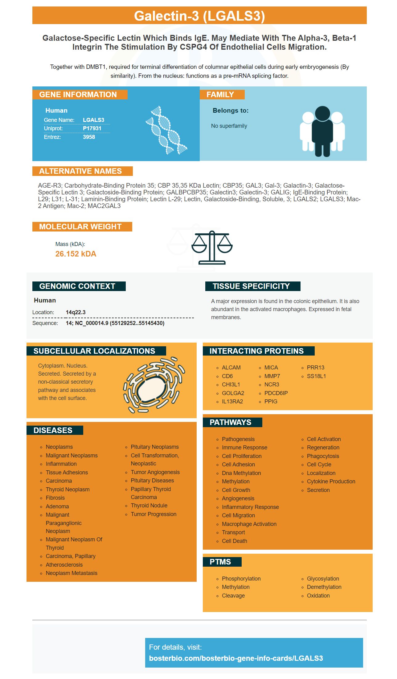
Facts about Galectin-3.
Together with DMBT1, required for terminal differentiation of columnar epithelial cells during early embryogenesis (By similarity). From the nucleus: functions as a pre-mRNA splicing factor.
Human | |
---|---|
Gene Name: | LGALS3 |
Uniprot: | P17931 |
Entrez: | 3958 |
Belongs to: |
---|
No superfamily |
AGE-R3; Carbohydrate-binding protein 35; CBP 35,35 kDa lectin; CBP35; GAL3; Gal-3; galactin-3; Galactose-specific lectin 3; Galactoside-binding protein; GALBPCBP35; Galectin3; Galectin-3; GALIG; IgE-binding protein; L29; L31; L-31; Laminin-binding protein; Lectin L-29; lectin, galactoside-binding, soluble, 3; LGALS2; LGALS3; Mac-2 antigen; Mac-2; MAC2GAL3
Mass (kDA):
26.152 kDA
Human | |
---|---|
Location: | 14q22.3 |
Sequence: | 14; NC_000014.9 (55129252..55145430) |
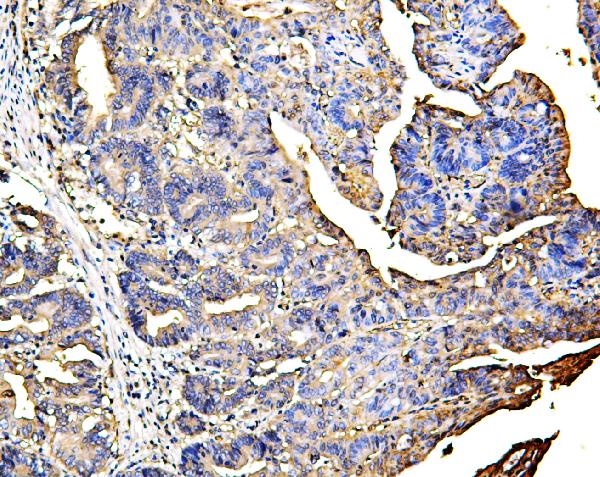
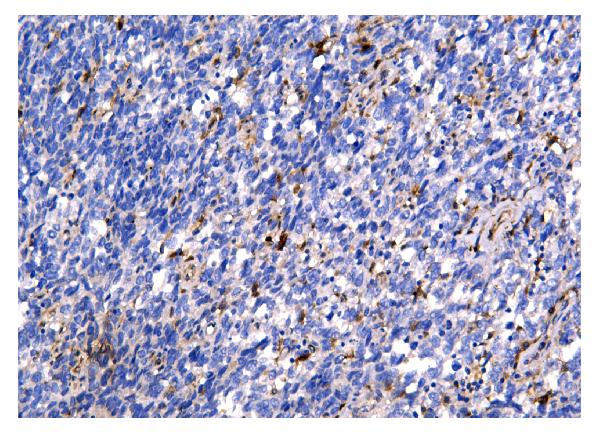
A major expression is found in the colonic epithelium. It is also abundant in the activated macrophages. Expressed in fetal membranes.
Cytoplasm. Nucleus. Secreted. Secreted by a non-classical secretory pathway and associates with the cell surface.
This article will examine the clinical applications of LGALS3 Marker. It is a molecule containing residues 117-244 along with regions 18-105. The following sections cover a few binding agents for the LGALS3 Marker. In addition, you'll be able to learn about MMP9 specific binding agents for the LGALS3 Marker.
A recent study suggests that LGALS3 is an essential player in managing the immune microenvironment in gliomas. Complex tumor microenvironments that surround gliomas include multiple cell types and a range of immune cell markers. One of these markers, CD163, targets M2 macrophages. These cells produce anti-inflammatory cytokines, encourage tumor growth and aid in growth. The identification of gliomas depends on the selection of suitable binding agents for LGALS3Marker.
LGALS3 is expressed in a variety of cell types. However, it is highly conserved throughout the brain. Its expression in the cerebellum is developmental regulated and coincides with the timing of excitatory synaptogenesis within the cerebellum. Similar to C1QL1, LGALS3 promotes neurite growth and neural cell adhesion. It also interacts in a functional way with integrins, who are crucial in synaptogenesis.
Although the exact role of LGALS3 is not yet known, recent studies have shown that it was a prominently expressed within the cerebellum during postnatal development. The marker is expressed in a subset of glial cells in the cerebellum, however it is dispensable for normal cytoarchitecture and excitatory synaptogenesis in Purkinje cells. It is also found in the fourth ventricle's choroid plexus.
A recent study revealed that LGALS3 protein expression is restricted to non-activated CD4+CD25+ cells. LGALS3 expression is highest in human CD4+CD25hi cells that have the FOXP3 signature. These studies revealed that UBD had no effect on LGALS3 expression. But LGALS3 mRNA expression in these cells was significantly increased similar to those of non-regulatory CD4+CD25+ cells that expressed FOXP3.
LGALS3 is a new gene that has the potential for clinical applications. It plays a significant role in neuronal movement and the guidance of axons. It also plays a role in regulating inflammation and neuronal apoptosis. In the cerebellar cortex LGALS3 is expressed in microglial cells. Additionally, it is involved in the formation of memory. We will now examine some of its clinical potential applications.
The expression of LGALS3 has been described in various pathological conditions in the mammalian brain, including reactive gliosis as well as oligodendrocytes. It has been observed in glial cells from different kinds in various parts of the brain, including the cerebellar and rostral migratory stream and olfactory bulbs. Certain studies have revealed that the protein can be located in neurons in the cerebellum as well as the hippocampus.
LGALS3 interacts with the cell adhesion molecule called integrin A3b1. This interaction results in the induction in vitro of LGALS3. Integrins play a significant role in synaptogenesis and neuronal development. It is located in the olivocerebellar system. Further, it is expressed in glial cells within the cerebellum. LGALS3 has clinical applications in brain imaging.
The role of the gene LGALS3 is vital to cerebellar development. Its expression is found in the cerebellar regions of mice who have been exposed to asphyxia at birth. Additionally, LGALS3 may also play a role in excitatory synaptogenesis within Purkinje cells. The gene LGALS3 could play a part in the assessment of brain ischemia as well as other neurodegenerative disorders.
Galectin-3 is encoded by the LGALS3 gene. It was initially proposed that this protein is responsible for the adhesion of macrophages, chemotaxis and the process of apoptosis. Its role in atherosclerosis composition and inflammatory profile were further studied. Thus, a search to find appropriate inhibitors of LGALS3 has been conducted. Two candidates were identified in this study that showed promising potential for activity against the gene.
Many computational methods have been employed to determine the affinity of molecules to their targets, including MMP9. One of these techniques is molecular docking which predicts how molecules will be directed to their targets. Molecular docking is a key instrument in drug design since it permits you to determine the affinities between molecules and their target proteins. This research focused on the search for and use of compounds that act as binding agents against activated MMP9.
The properties of these compounds make them suitable to conduct studies that inhibit. TIMP-1 has been shown to be inhibited by various proteins. In vitro assays, the use of thiol-modifying agents, such as HgCl2, 4-aminophenylmercuric acetate, and N-ethylmaleimide, is a useful method to stimulate MMP activity.
A homotrimeric structure of MMP-9 has been suggested. AFM and TEM tests revealed that the trimer was unstable in reducing conditions. This indicates the involvement of disulphide bridging. Two distinct populations were identified using molecular imaging measurements. They had an identical overall structure but different dimensions. Images from molecular imaging showed that the smaller complexes are the most abundant. The inner diameters are determined by the absence of subunits in between them, while the outer diameters represent the length of subunits.
The results of the current study show that ligands that attach to the catalytic ion of MMP-9 have a high affinity and specificity for the catalytic domain. These findings will be useful in the design of low-molecular-weight compounds that are highly dependent on MMP-9. One of the difficulties is identifying these compounds, however, since they tend to be difficult to obtain and unstable.
MMP-2, MMP-9, as well as collagen binding agents were identified and the results were compared. These activities were linked to age and pathophysiology, as well as the discovery of new markers of longevity. A Sicilian study found that MMP-2 activity levels were correlated with haematochemical parameters that could be used to predict longevity. These findings demonstrate the need to have an agent that binds to these enzymes.
PMID: 2261464 by Robertson M.W., et al. Human IgE-binding protein: a soluble lectin exhibiting a highly conserved interspecies sequence and differential recognition of IgE glycoforms.
PMID: 2402511 by Cherayil B., et al. Molecular cloning of a human macrophage lectin specific for galactose.
*More publications can be found for each product on its corresponding product page