This website uses cookies to ensure you get the best experience on our website.
- Table of Contents
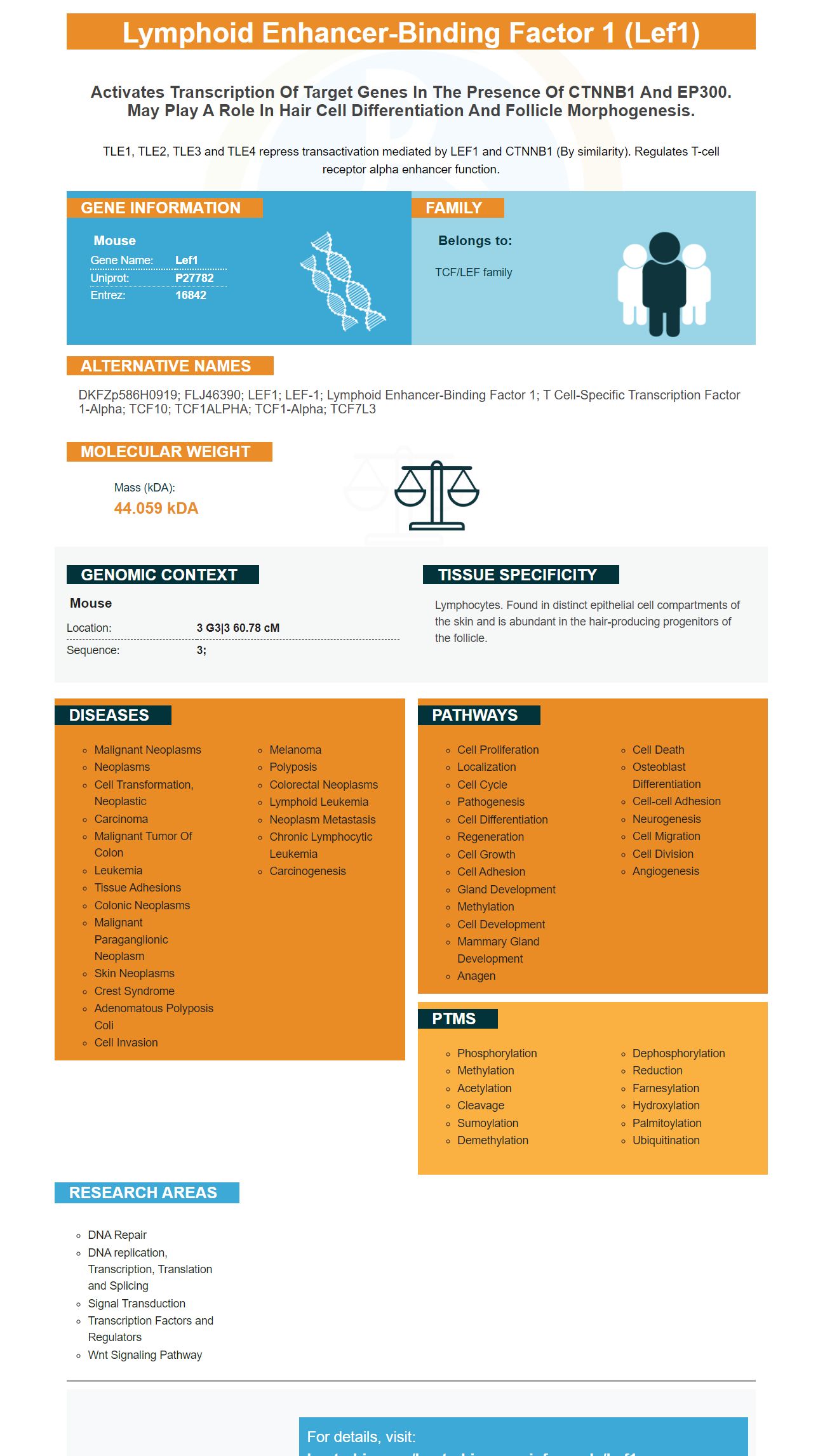
Facts about Lymphoid enhancer-binding factor 1.
TLE1, TLE2, TLE3 and TLE4 repress transactivation mediated by LEF1 and CTNNB1 (By similarity). Regulates T-cell receptor alpha enhancer function.
Mouse | |
---|---|
Gene Name: | Lef1 |
Uniprot: | P27782 |
Entrez: | 16842 |
Belongs to: |
---|
TCF/LEF family |
DKFZp586H0919; FLJ46390; LEF1; LEF-1; lymphoid enhancer-binding factor 1; T cell-specific transcription factor 1-alpha; TCF10; TCF1ALPHA; TCF1-alpha; TCF7L3
Mass (kDA):
44.059 kDA
Mouse | |
---|---|
Location: | 3 G3|3 60.78 cM |
Sequence: | 3; |
Lymphocytes. Found in distinct epithelial cell compartments of the skin and is abundant in the hair-producing progenitors of the follicle.
You've come to a good location if you're thinking about LEF1 as a treatment option for cancer. This biomarker inhibits E–cadherin's production and also promotes expression SNAI2 (Slug). Find out more about its applications in cancer research. This biomarker is a natural antagonist of E-cadherin and has a number of uses in cancer research.
This gene is upregulated in some types of glioma tissue . It is an excellent biomarker to detect the presence of this type of tumor. This gene is also present in non-tumor tissue. Its function is not completely understood. Boster Bio's LEF1 test can be used to determine LEF1-AS1 expression. LEF1-AS1 knockdown in glioma tissues inhibits the growth of tumor cells.
LEF1 is a functional protein that carries out a variety of biological functions in the body. It has been linked to multiple kinds of cancer. It has been found to play a key role in glioma. It also inhibits the expression of miR-218, which is a tumor promoter. It has been shown to be involved in prostate cancer.
LEF1 was identified as being linked to FN1 in research. Inhibition of this gene leads to the expression of FN1. MicroRNAs could also regulate it. LEF1 knockdown reduced the SNAI2 reporter's activity. Additionally, SNAI1 expression was found to increase in depleted LEF1 U2OS cells. This study confirms that LEF1 plays a role in activating FN1 transcription.
LEF1 is involved in the regulation of mesenchymal cell migration. LEF1 is knocked out to facilitate the recruitment of CDh2 cell-cell contacts. F-actin remains intact. Western blotting was employed for measuring the levels of CDh2 protein and LEF1 protein with their control. The CDh2 levels were measured relative to VCL which was used as a loading control. qRTPCR was also used to determine the mRNA levels for CDh2 (and FN1) using the qRTPCR. ELISA was used to measure LEF1 and CDh2 levels in response to LEF1 knockdown.
Autophagy plays an important role in maintaining epithelial characteristics. Inhibiting autophagy can decrease the levels of E. cadherin protein and could cause changes in the cell-cell junction. Here are the possible targets of Boster Bio LEF1 inhibitors to E-cadherin. These compounds have demonstrated promising results in cell research.
The saRNA-mediated activation of E-cadherin expression in BCa cells inhibits their growth and metastasis. These compounds also reduce expression of b-catenin/TCF target genes. Boster Bio LEF1 inhibitors can have a positive effect on BCa tumors, according to the findings. The saRNA-mediated activation of E.cadherin expression inhibits growth and metastasis of BCa cancer cells.
The deficiency in E-cadherin's regulation has been linked to poor prognosis for patients and invasiveness of tumours. The adhesion protein is present in cells. It also interacts with b-catenin, which translocates to the nucleus and activates several promigratory and epithelial-mesenchymal transition genes. Therefore, aiming to reduce the activity of E-cadherin through an inhibitor of LEF1 is a feasible approach to improving the outcomes of patients.
In cells that express E'cadherin, LC3B expression increased. In addition, E-cadherin and the LC3B colocalization was increased. These results suggest that Boster Bio LEF1 inhibits could improve the treatment of MDAMB-231 cancer cells. This study demonstrates the potential of LEF1 inhibitors for cancer treatment. These compounds can also be used to prevent cancer growth.
SNAI2 is a cell migration factor that plays an important role in the early development of the neural tube. It is thought to function by promoting epithelial-mesenchymal transition, an important process that regulates cellular migration, survival, and other processes. However, its precise role in the development of breast cancer cells is not known. This study offers additional information on how SNAI2 is able to facilitate the growth and migration of breast cancer cells.
In a previous study we discovered that the overexpression the LEF1 gene decreased SNAI2 expression. The overexpression of miR-452 slowed the VEGFA-induced increase in invasion. This suggests that miR-452 blocks Slug expression which is essential for cell mobility. Our results suggest that the LEF1 gene is critical for the induction of SNAI2.
Moreover, miR-452 regulates SNAI2 expression through Sox2. The miR-452 targets the 3''UTR on the SNAI2. We performed a miRNA screen to identify miRs which target SNAI2 cells by using MDA-MB-231 cells. TargetScan was used to study 47 miRNA expressions. Interestingly, miR-452 had a high probability score of targeting SNAI2 that microT-CDS confirmed.
TGF-b3 neutralizing antibody blocked the expression of E-cadherin vimentin and fibronectin following Snail and Slug treatments in MDCKII cells. The results of immunoblotting indicated that these proteins impeded movement and invasion triggered by Snail activity. Similar results were observed when we employed pcDNA3E expression vectors in MDCKII cells and A375 cells.
A number of studies have shown that LEF1 expression is associated with poor prognosis. The study in this study showed that nuclear LEF1 expression was detected in nine of the 25 samples (36 percent) of patients suffering from brain metastases due to Adenocarcinoma. TCF4 was present in all samples. Nuclear LEF1 expression was only found in three of nine Grade 1+ samples. All patient samples included the LEF1 marker which was also expressed along with b-catenin. However, the connection between the two genes is weak.
Although LEF1 expression was found to be a poor prognostic factor in adult patients newly diagnosed with AML however, it is an intriguing candidate gene to be used for risk stratification. The results also suggest that LEF1 could be a target for therapy. However, the exact role of LEF1 in MM is not yet known. The study also revealed an increased risk of disease progression in patients who have low LEF1 expression.
In a different study, LEF1 expression was linked to lower overall survival for patients with high levels of LEF1 expression. LEF1 expression was also related to a lower median age. LEF1 expression was significantly different between patients with advanced age and those who were 60 years old. The findings of this study have implications for the treatment of patients suffering from cancer. If it is a prognostic factor, the next step is to evaluate whether it will improve the quality of life.
The LEF1 gene encodes a protein that serves multiple functions, including controlling cell growth, death of cells, tumor immunity, and controlling cellular proliferation. Its best known function is to bind an antigen that is associated with tumors and initiate an immune response. Other possible functions include anchoring additional transcription factors as well as facilitating the activation of immune cells. It also is involved in stem-like cell growth. There are numerous known antigens for tumors that target the gene that are involved in cancer, for example.
Nelfb is a key factor in promoting chromatin access. The number of CD8+ human primary CD8+ cells with high levels of this gene increased by overexpression. This implies that NELF could be the rate-limiting factor for T cells of the CD8+ family that have antitumor activity. Additionally, it is thought to boost memory and the polyfunctionality of T cells.
PMID: 1827423 by Travis A., et al. LEF-1, a gene encoding a lymphoid-specific protein with an HMG domain, regulates T-cell receptor alpha enhancer function.
PMID: 8415007 by Fujimoto S., et al. Nucleotide sequence of a cDNA encoding an alternative form of LEF- 1.