This website uses cookies to ensure you get the best experience on our website.
- Table of Contents
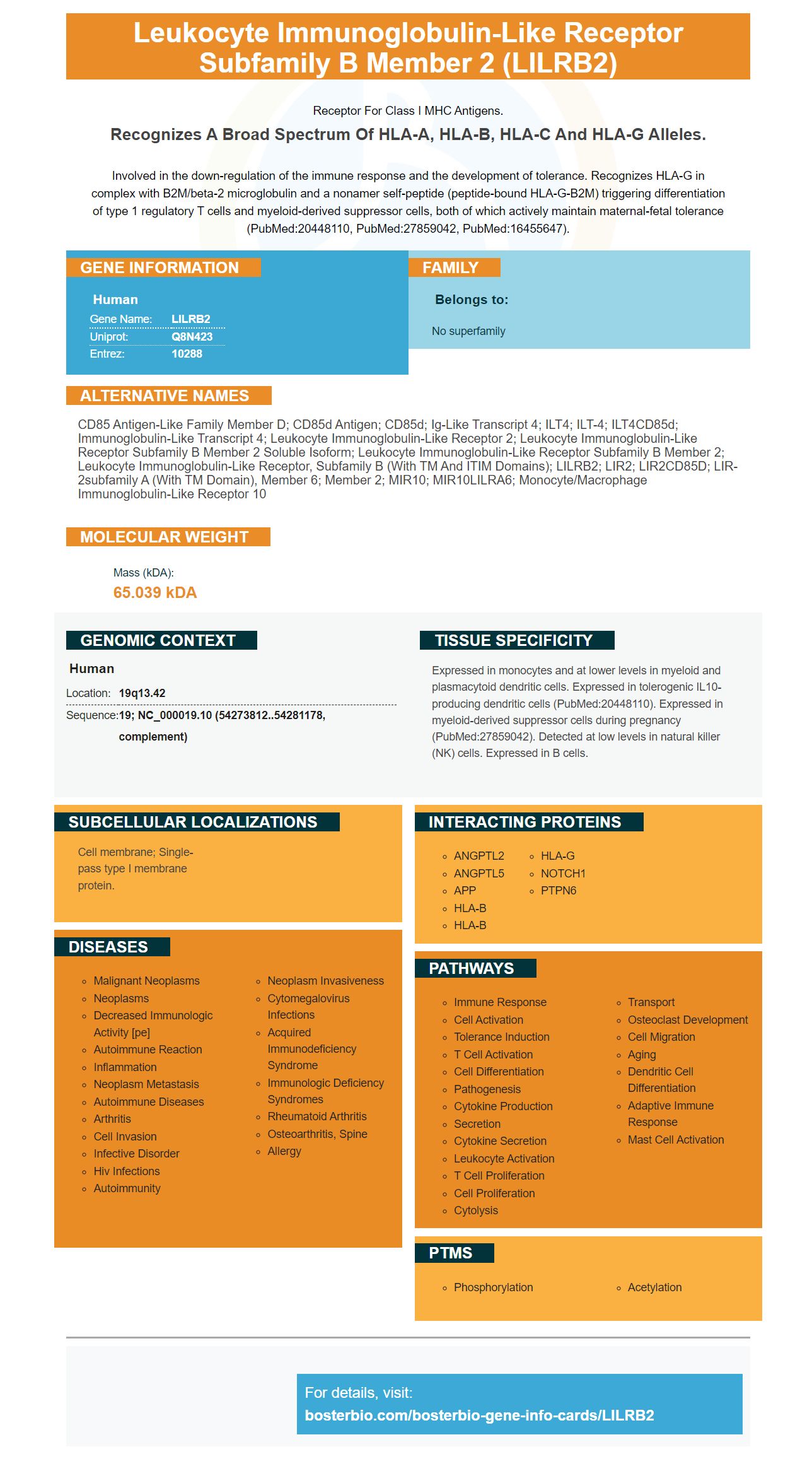
Facts about Leukocyte immunoglobulin-like receptor subfamily B member 2.
Receptor for class I MHC antigens.
Recognizes a broad spectrum of HLA-A, HLA-B, HLA-C and HLA-G alleles.Involved in the down-regulation of the immune response and the development of tolerance. Recognizes HLA-G in complex with B2M/beta-2 microglobulin and a nonamer self-peptide (peptide-bound HLA-G-B2M) triggering differentiation of type 1 regulatory T cells and myeloid-derived suppressor cells, both of which actively maintain maternal-fetal tolerance (PubMed:20448110, PubMed:27859042, PubMed:16455647).
Human | |
---|---|
Gene Name: | LILRB2 |
Uniprot: | Q8N423 |
Entrez: | 10288 |
Belongs to: |
---|
No superfamily |
CD85 antigen-like family member D; CD85d antigen; CD85d; Ig-like transcript 4; ILT4; ILT-4; ILT4CD85d; Immunoglobulin-like transcript 4; Leukocyte immunoglobulin-like receptor 2; leukocyte immunoglobulin-like receptor subfamily B member 2 soluble isoform; leukocyte immunoglobulin-like receptor subfamily B member 2; leukocyte immunoglobulin-like receptor, subfamily B (with TM and ITIM domains); LILRB2; LIR2; LIR2CD85D; LIR-2subfamily A (with TM domain), member 6; member 2; MIR10; MIR10LILRA6; Monocyte/macrophage immunoglobulin-like receptor 10
Mass (kDA):
65.039 kDA
Human | |
---|---|
Location: | 19q13.42 |
Sequence: | 19; NC_000019.10 (54273812..54281178, complement) |
Expressed in monocytes and at lower levels in myeloid and plasmacytoid dendritic cells. Expressed in tolerogenic IL10-producing dendritic cells (PubMed:20448110). Expressed in myeloid-derived suppressor cells during pregnancy (PubMed:27859042). Detected at low levels in natural killer (NK) cells. Expressed in B cells.
Cell membrane; Single-pass type I membrane protein.
The LILRB2 marker has been identified as a promising marker in the diagnosis of human acute leukemia (AML) and human monocytic AML cells. Moreover, it shows better overall survival in AML patients than CD14. But why is LILRB2 a superior marker? Here are the possible explanations. Find out more.
In the study presented here, LILRB2 expression was found to be higher in the leukocyte subsets of the mAb-treated AML cells than in control human blood. The expression level of LILRB2 was assessed using one-way ANOVA, which used five independent donors. LILRB2 expression was more prominent in the CD8a gene which regulates the cytoskeletal structure.
The researchers also identified the cytotoxic activity of CAR-T cells directed against anti-LILRB4 against multiple types of AML cell lines. After co-culture for 24 hours with anti LILRB4 CAR–T cells, the anti LILRB4 cell-t cells showed significant cytotoxicity towards leukemia cells. These CAR-T cells also showed significant toxicities against the AML cell line as well as normal LILRB4+ primary monoocytes.
LILRB3 ligation regulates the activation and proliferation of T cells. LILRB3 expression is found in human monocytes. LILRB3 Abs do not inhibit the proliferation of T cells in the absence monocytes. This finding is supported in part by binding epitopes to mAbs that can modify the receptor.
LILRB2 expression was also more common in human Leukemia than CD14. The expression of LILRB4 on human leukemia cells was also significantly higher than that of CD14. LILRB4 expression was also linked to the presence of CD34 which is a marker for leukemia stem cells. In addition, this protein was found to be co-expressed with CD34 and CD45RA, which were previously considered non-specific markers for human leukemia.
This result is consistent with previous findings. LILRB2 is more sensitive to LILRB4 than CD14, which may allow the cancer cell to escape the immune reaction. LILRB4 could also help tumor cells to evade the immune system. Further research is needed to determine if LILRB2 is a better indicator than CD14.
The study used an LILRB3-specific phage display library as a tool to evaluate the binding properties. The generated protein was immobilized and various mAbs were flown across the chip. The mAbs reacted cross-reactively with CHO-S cells in order to identify LILRB3 positive target-transfected cells. These clones were then tested for specificity using ELISA.
LILRB2 is more sensitive to AML than CD14. In fact, LILRB2 has more sensitivity than CD14 in the detection of differentiated leukemia cells. LILRB4 is less sensitive than CD14 and therefore harder to detect. Mutagenic primers can increase its expression.
The mAbs that were used in this study were created to identify cytoplasmic/nuclear antigens of CD14+/CD16+ ukemia cells. The experiment used several combinations of fluorescent reagents. To detect a subset leukemia cells, mAbs against CD34, MPO and CD7 were used.
In the binding assay, h228-3 antibody binds the D1 domain to the cytoplasmic-hemocyte antigen. Fig. 2E shows the binding surface of antibody to D1. 2E. 2E. LILRB2 also binds LILRB4ECD, a mAb more effective than CD14 at detecting differentiated acute lymphoma cells.
In a similar study, LILRB2 was also a better marker for identifying acute leukemia cells than CD14. These results confirm previous findings that LILRB2 was more sensitive than CD14 in distinguishing AML-cells. Moreover, the increased sensitivity of LILRB2 may mean a higher rate of remission in AML patients.
LILRB2 shares a structure with CD14 when it is molecularly modelled. LILRB2 contains a BC loop which interacts with h228-3, and binds to CD14. LILRB4 has both the BC loop and the h228-3 binding motif, which is why LILRB2 is better than CD14 in differentiated acute leukemia cells.
LILRB4 has anti-AML capabilities. LILRB4 can be activated by ADCC-mediated AML cell destruction using monoclonal humanized antibodies. This makes LILRB4 attractive as an anti-AML drug target. However, this strategy must be validated in order to be effective in treating AML.
A higher overall survival rate for patients with AML is associated to the LILRB2 genetic. This gene plays dual roles in tumor biology. It acts as both immune checkpoint molecules, and tumor-sustaining factor. LILRBs also did not alter normal development in knockout mice. They could be considered ideal targets for treatment due to their dual role in tumor biology. This review summarizes our knowledge on the expression patterns, ligands, signaling pathways, and functions of the LILRB family members.
Another measure of overall longevity is the five-year survival ratio (RSS). It shows how many patients survive five years after treatment. It is calculated for patients over 20 years old. It is difficult, however, to predict the survival rate in younger patients. The five-year survival ratio varies from patient a patient and is dependent upon several biological features. A majority of patients with AML will survive the treatment. This includes chemotherapy and bone Marrow transplant.
In addition, the LILRB2 gene family is associated with increased levels of immune cells in AML patients. The LILRB2 genes were linked with both DSS AND OS. This suggests LILRB2 could be an immune inhibitory regulator that aids infiltrating tumors. These results support the notion that LILRB2 is a more accurate marker than CD14 for predicting overall survival in human AML patients.
LILRB2 is a better marker for human AML than CD14. Besides, LILRB2 is more sensitive than CD14 for predicting overall survival in AML patients. The study authors thank the patients for their consent and the Institute of Hematology and Blood Transfusion in Prague for their participation in the study.
LILRB2 also predicts better overall survival rates in AML than the DfN and LAIP trials. The clinical benefit of LILRB2 remains unclear. There are numerous unpublished reports suggesting that the DfN has lower sensitivity than the LAIP. The expression of NPM1 is variable, making it difficult to predict MRD's outcome. Patients without a comprehensive antibody panel may experience false negatives.
LILRB2, a transmembrane type of glycoprotein that contains extracellular Ig -like domains, is known as human leukocyte immuneglobulinlike receptor-like receptor B2. The LILRB2 family binds to cytokines, T cell antigens, and peptides. These antigens can be found on many types of leukemia cells including acute myeloid (AML) and cytokines.
LILRB2 inhibition inhibited leukemia development on mouse xenografts. Activation of the marker inhibited the immune system. These results are consistent in line with the Boster Bio study. Inhibition of LILRB2 can also inhibit the immune system. LILRB4 may, however.
The anti-LILRB4 antibodies have been shown to block the activation of human LILRB4 by APOE, a functional ligand. Antibodies bind LILRB4 (and APOE) in the ELISA. Mouse serum is a positive test. A murine model of AML was also used to test for antibodies to LILRB4.
Boster Bio used anti-LILRB4 antibodies to test the antibodies' ability suppress leukemia in mice. The mice were given anti-LILRB4 antibodies on day 0, day 3, and day 6, respectively, after the transplant. After the administration of antibodies, mice were sacrificed. The flow cytometry analysis of their livers and bone marrow was used to analyse their peripheral blood. Both mouse models showed significant inhibition in the development of AML by anti-LILRB4 antibodies.
LILRB family members are highly expressed in leukemia cells. They correlate negatively with overall survival rates in patients with AML. Many LILRBs are necessary for the survival and development of human leukemia stem cell cells. Because they are required for leukemic stem cells, inhibiting their activity in the immune system indirectly enhances anti-tumor effects. Researchers are looking into LILRB signaling to treat AML.
The antigen-binding proteins are useful fragments of antibodies that bind a specific target antigen. These fragments include CDRs (antibodies), and variable domains (heavy and light) of the heavy and medium chains. The variable regions of these chains are typical locations for antigen-binding site. They also form the antigen binding sites. This fragment is useless by itself because it lacks a natural ligand.
PMID: 9382880 by Colonna M., et al. A common inhibitory receptor for major histocompatibility complex class I molecules on human lymphoid and myelomonocytic cells.
PMID: 9548455 by Borges L., et al. A family of human lymphoid and myeloid Ig-like receptors, some of which bind to MHC class I molecules.