This website uses cookies to ensure you get the best experience on our website.
- Table of Contents
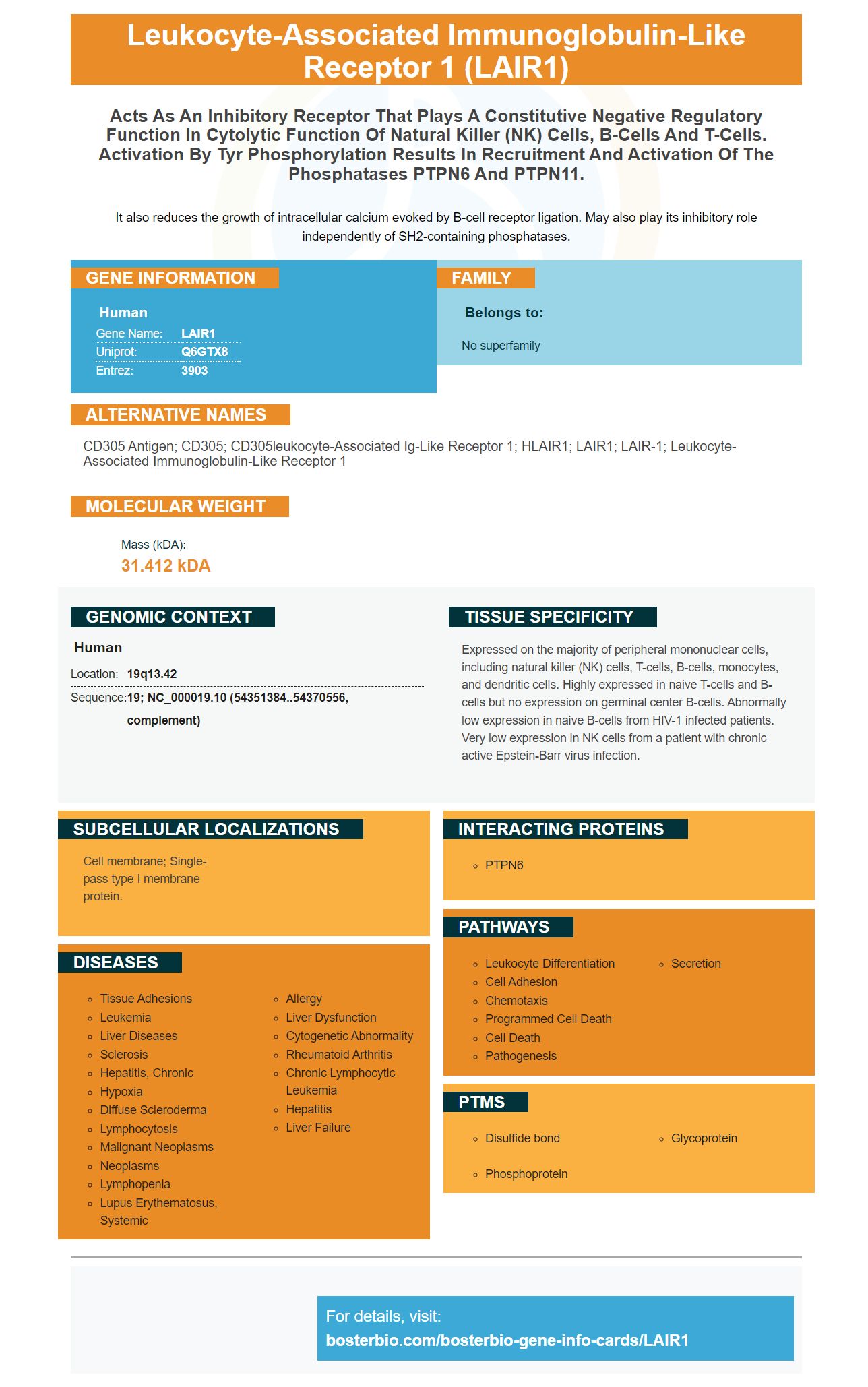
Facts about Leukocyte-associated immunoglobulin-like receptor 1.
It also reduces the growth of intracellular calcium evoked by B-cell receptor ligation. May also play its inhibitory role independently of SH2-containing phosphatases.
Human | |
---|---|
Gene Name: | LAIR1 |
Uniprot: | Q6GTX8 |
Entrez: | 3903 |
Belongs to: |
---|
No superfamily |
CD305 antigen; CD305; CD305leukocyte-associated Ig-like receptor 1; HLAIR1; LAIR1; LAIR-1; leukocyte-associated immunoglobulin-like receptor 1
Mass (kDA):
31.412 kDA
Human | |
---|---|
Location: | 19q13.42 |
Sequence: | 19; NC_000019.10 (54351384..54370556, complement) |
Expressed on the majority of peripheral mononuclear cells, including natural killer (NK) cells, T-cells, B-cells, monocytes, and dendritic cells. Highly expressed in naive T-cells and B-cells but no expression on germinal center B-cells. Abnormally low expression in naive B-cells from HIV-1 infected patients. Very low expression in NK cells from a patient with chronic active Epstein-Barr virus infection.
Cell membrane; Single-pass type I membrane protein.
The LAIR1 marker is a great option for research in many fields. This monoclonal antibody contains antigen-binding elements. Its primary use is in immunohistochemistry. Monoclonal antibodies are proteins that have the same molecular weight and affinity as the target antigen. They are often used in cell cultures as reagents.
Figure 1 shows the structure of an anti-inflammatory antibody containing the LAIR1 mark. Figure 1 shows the structure and antigen-binding fragments of LAIR1 antibodies. LAIR1 insert, in contrast, is oriented along the long axis. It interacts seamlessly with all five CDR loops. This structural arrangement can only be achieved by long linkers.
Monoclonal antibodies that bind LAIR1 are characterized by their high specificity, with a KD value of ten-6 M or less. When equilibrium is achieved, the antigen-binding ability is measured by the dissociation and the association rates, or koff/kon. It can be expressed using koff/kon. This is a rate at which monoclonal antibodies are able to bind a specific antibody.
Human HEK293T cells demonstrated that C1q, which is a protein inhibitor for TLR signaling has inhibited anti-LAIR-1 activities. Interestingly, LAIR-1 and C1q share the same functions in the immune system. Both proteins have collagen-like tails that are involved in suppressing immune cell activity. C1q can also inhibit monocyte derived DC differentiation and prevent pDCs producing IFN-1a.
Three distinct mutations in the LAIR1 gene affect the Fab's ability to bind collagen. A significant portion of LAIR1-containing antibody sequences contains mutations in R59 (E61) and R65 that affect collagen binding. However, mutations in RIFIN binding are less important than those in LAIR1.
Furthermore, mice lack a LAIR-2 homolog, suggesting that interaction between C1q & LAIR-1 is not dependent upon layered regulation. LAIR-1-deficient mice made more IFNa than wild-type mice in vivo. These preliminary findings suggest that LAIR-1 and IFN-a interact in vivo. However their exact roles are not known.
The design of new therapeutic antibodies will be possible thanks to the discovery of LAIR1 antigen binding peptide. In fact, this type of fusion protein may be used as a marker to help identify cancer cells. However, this approach is not without its limitations. This mechanism has potential to be used in therapeutic development. It has many uses and can lead into new discoveries.
LAIR-1 is different from LAIR-2 in terms of binding kinetics. But these antibodies are capable of detecting purified proteins by using a solid-phase-binding microassay. They can also detect C1q collax tails. LAIR-1 is a better C1q collagen binding partner than LAIR-2. A comparison between these two types of antibodies can help determine if a patient has cancer or other health-related factors.
Five CDR loops are found in the antigen-binding fragment Lair1 gene. One of these loops is partially blocked by LAIR1. The LAIR1-inserted CDRs interact directly with the two CDRs in the heavy chain's first two CDRs. The two CDR loops of the light chain interact with one of the two linkers, the C93 of the heavy chain and C223 of the linker 2 of the light chain form a disulphide bond.
Monocytes express LAIR1 marker. This molecule is responsible to the upregulation LAIR1 in the inflammatory phase. Interestingly, LAIR1 expression is restored to normal levels during the resolution phase. Its upregulation has been associated with inflammation, such as in the cases of sepsis monoocytes, rheumatoid arthritis, and liver cirrhosis. TLR signaling and IFN signaling, which are both inflammatory responses, also increase LAIR-1 expression.
The structure of LAIR1 marker was revealed. The Fab, or 'L, chain, is composed two helices, which are linked by a sulphide bond. This structure provides a clearer view of how the antibody recognizes the LAIR1 ligand. It is not known if this linkage has a rigid or flexible structure. A group of researchers recently validated monoclonal antibodies against the LAIR1 marker.
The LAIR1 molecule has a kinked CDR H3 loop in the heavy chain. It spreads to both LAIR1 termini when it binds with the Fab. However, the LAIR1 marker was not fully studied by Winn et al. This discovery is important because it shows how LAIR1 affects the accessibility of CDR loops. The AREAIMOL marker can also be used for this purpose but it is not yet available in monoclonal antibodies against the LAIR1 mark.
LAIR-1, a classical dendritic cell subpopulation, has been found to be highly expressed on monocytes. Differentiated monocytes (CD1c+ CDDC2 and CD141+ CDDC1 cells) have different levels and phenotypic characteristics. They also all have high levels in cytokines.
In 2016, a new class was discovered of antibodies that contained the entire human-derived protein embedded into the CDR Loop. These antibodies bind directly to the RIFIN molecule in human red blood cell cells. They are also infected from the malaria parasite. It is unclear how these antibodies bind RIFINs. Further studies are needed to determine how these antibodies function.
The LAIR1-binding Lair1 antibodies can enhance antigen-specific responses in the patient. They can be used to treat autoimmune and inflammatory diseases. Targeting extrafollicular immune responses may be beneficial for treating these diseases as GCs are thought of being hotbeds to B cell development. Monoclonal antibodies that are specific for the LAIR1 marker have been developed. This is a promising start.
It is still not known what role autoantibodies may play in MS's pathogenesis. Although some studies have shown that B cells may play a role in MS, others have shown that these antibodies are not responsible for the disease. Although this is a good step forward, more research is needed to understand the mechanism behind this autoantibody. Monoclonal antibodies against LAIR1 only work in a small number of patients.
PMID: 9285412 by Meyaard L., et al. LAIR-1, a novel inhibitory receptor expressed on human mononuclear leukocytes.
PMID: 10229813 by Meyaard L., et al. Leukocyte-associated Ig-like receptor-1 functions as an inhibitory receptor on cytotoxic T cells.