This website uses cookies to ensure you get the best experience on our website.
- Table of Contents
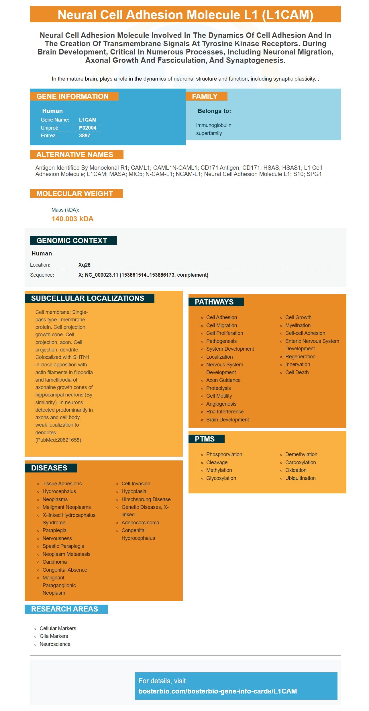
Facts about Neural cell adhesion molecule L1.
In the mature brain, plays a role in the dynamics of neuronal structure and function, including synaptic plasticity. .
Human | |
---|---|
Gene Name: | L1CAM |
Uniprot: | P32004 |
Entrez: | 3897 |
Belongs to: |
---|
immunoglobulin superfamily |
antigen identified by monoclonal R1; CAML1; CAML1N-CAML1; CD171 antigen; CD171; HSAS; HSAS1; L1 cell adhesion molecule; L1CAM; MASA; MIC5; N-CAM-L1; NCAM-L1; neural cell adhesion molecule L1; S10; SPG1
Mass (kDA):
140.003 kDA
Human | |
---|---|
Location: | Xq28 |
Sequence: | X; NC_000023.11 (153861514..153886173, complement) |
Cell membrane; Single-pass type I membrane protein. Cell projection, growth cone. Cell projection, axon. Cell projection, dendrite. Colocalized with SHTN1 in close apposition with actin filaments in filopodia and lamellipodia of axonalne growth cones of hippocampal neurons (By similarity). In neurons, detected predominantly in axons and cell body, weak localization to dendrites (PubMed:20621658).
In this article, we'll discuss the advantages of using the L1CAM marker in biomedical applications, the best way to properly prepare samples for this purpose, and what methods are available to use the L1CAM marker. This information is beneficial to biomedical researchers around the globe. Boster provides product credits to those who publish their results using the L1CAM marker. How can scientists get started using the L1CAM marker
One of the most frequent questions doctors ask is whether or whether the L1CAM marker is useful in the detection of endometrial cancer. Although the overall rate of recurrence is low, patients with tumors express L1CAM were at greater risk of recurrence. While L1CAM positivity was associated with a lower overall survival however, it wasn't associated with a lower survival rate for patients free of disease.
L1CAM is a transmembrane-specific protein that is found primarily in GABAergic interneurons and parvalbumin+ within adult cerebral cortex. Munakata and coworkers. L1CAM is a potential anticancer therapy, specifically targets tumor-promoting cell.
The positive results of L1CAM staining were assessed by two independent pathologists blinded to the clinical information of the patients. The specimens were graded based on their intensity. A negative control was constructed by replacing the primary antibody with PBS. Only specimens with high intensities were considered to be positive for L1CAM expression. To determine low or no L1CAM expression to determine whether L1CAM expression was low or not, the ROC curve cut-offs were used.
In addition to its prognostic value, L1CAM expression is a viable therapeutic target for high-risk endometrial cancer. A threshold greater than 50% can be a predictor of prognosis in high-risk carcinoma. Although p53 and L1CAM have a mechanistic association, L1CAM expression could be an unidentified group of non-endometrioid differentiated cells.
The detection methods of the L1cam marker are typically performed with immunohistochemistry (IHC) on formalin-fixed paraffin-embedded tissues, which are easily accessible for routine storage of tissues. Although IHC is a semiquantitative method however, its accuracy is not high because of the inherent bias of observers. IHC must be conducted with precision.
L1CAM is abundant on the cell surface of replicative senescent fibroblasts. This is due to up-regulation of cell-surface proteins in these cells. The protein fractions were collected with streptavidin columns, then validated using Western Blotting. In addition to Western blotting, FACS analysis of BJ fibroblasts revealed the presence L1CAM on their surface.
The diagnostic accuracy of an L1CAM test can be evaluated by using its predictive value and likelihood ratios. This marker should be identified accurately using a reliable method. Once the L1CAM assay has been validated and confirmed, it is time to design an accurate method. The most appropriate method for a given patient's case is one that is highly reproducible. If the L1CAM test is not successful then the patient should be immediately referred to a doctor.
A single antibody against L1CAM is highly specific. In one study Anti-L1CAM antibodies have been co-coated with magnetic beads. These beads enabled specific immunocapture of neuronally derived exosomes. Simple sensor surfaces can be used to increase capture specificity. The statistically significant distinction of PD from control samples was made possible because of the robustness of the internal markers. This novel method for identifying PD exosomes has a myriad of promising applications.
Recent research has proven that L1CAM expression is linked to lymph node metastasis EJA or GC. The study also demonstrated that L1CAM plays an important role in lymph node metastasis, despite its low expression in lymph nodes. In the same way, L1CAM is part of the cell adhesion molecules family that may promote endothelial cell migration in cancer cells through a receptor-ligand interaction.
A magnetic beads coated with anti-L1CAM (MB) are utilized to identify an internal protein marker. A reagent-free impedimetric assay (RES) can be used to determine the content of internal exosome markers, such as a-synuclein and syntenin-1. The quantitative analysis of exosomes could be used to determine significant differences between PD patients and healthy controls.
A novel variant of L1CAM was discovered in mice. It binds to a protein called L1CAM which normally joins adjacent cell. The researchers then targeted this protein using an chimeric protein fusion called NOVA2. This modified L1CAM helps to promote the development and growth of new blood vessels. This way, the L1CAM marker could have multiple applications. It can also be used to detect ovarian cancer.
The L1CAM gene is found in human normal tissues like brain, ovary, heart, the trachea, and liver. To analyse cancerous ovarian tissue, RT-PCR is carried out using the primers hL1E24_F and hL1E26_R. Pathologists must select the appropriate areas to analyze the cancerous tissue of the ovarian and prepare the samples.
In mouse and human cells NOVA2 has been identified as a regulator of L1CAM splicing. This gene also plays the capacity to regulate L1CAM expression. In addition to regulating L1CAM expression, NOVA2 also triggers exon skipping and inclusion when it is bound to the upper intronic region. Both human and mouse L1CAM exons are enriched with YCAY repeats.
L1CAM is also a versatile protein with a variety of clinical applications. It regulates cell-cell adhesion and is an anti-angiogenic protein. L1CAM interacts with many membrane proteins in addition to its anti-angiogenic properties. These interactions have made it possible to enhance L1CAM detection in many medical fields. The L1CAM gene is cloned by using a specific DNA sequence and cell type.
In this study, tumors that showed high levels of L1CAM expression were at a higher risk of developing metastasis when they are detected in an early stage of disease. Patients with high L1CAM expression are also more prone for metachronous metastasis. This is that the majority of deaths due to cancer in western countries take place. Despite recent advancements in multimodal therapies the colorectal cancer remains a major cause for cancer-related morbidity. Within three years of diagnosis metachronous metastases occur in 86 percent of patients.
This study shows that L1CAM is associated with the tumor-stroma interface. E-cadherin, slug and the L1CAM were both negatively associated. Additionally, L1CAM was also correlated with cytosolic beta-catenin, which indicates that tumors are infected with tumor cells that express this protein. Consequently, L1CAM is an extremely sensitive marker for detecting cancer-related diseases.
Using SIMOA assay, researchers have been measuring the vesicle markers in human plasma and CSF. The markers L1CAM and membrane tetraspanins were discovered to be eluted using albumin that is soluble and smaller, lighter structures, suggesting that they are abundant in extracellular vesicles. These findings are crucial in diagnosing vascular diseases and treating strokes that are ischemic.
L1CAM is a distinct molecule of neural cells and has been discovered to be expressed in 97% of gliomas as well as Astrocytomas. L1CAM can be distinguished from a paraganglioma without IHC. However, L1CAM can be used as a standard marker for both kinds of tumors. The technical details of the IHC test could affect the performance of L1CAM in SPN vs. the NET differentiating diagnosis.
The cell structure of the L1CAM marker is identical in both genders. L1CAM is found in adults in the urethra, as well as in the anorectal canal. L1CAM mRNA can be found in the womb and ectocervix of the fetus. L1CAM expression in these tissues could signal an illness, since it is found in both foetuses.
The L1CAM marker can aid in identifying mutations in L1CAM genes in the fetal brain. Hydrocephalus is diagnosed with L1CAM gene mutations in the fetal brain tissue. There are a variety of methods to conduct the test and the sample preparation is based on the particular kind of fetal tissue. One of the most popular methods is the Western blot.
PMID: 1932117 by Kobayashi M., et al. Molecular cloning of cell adhesion molecule L1 from human nervous tissue: a comparison of the primary sequences of L1 molecules of different origin.
PMID: 1769655 by Hlavin M.L., et al. Molecular structure and functional testing of human L1CAM: an interspecies comparison.