This website uses cookies to ensure you get the best experience on our website.
- Table of Contents
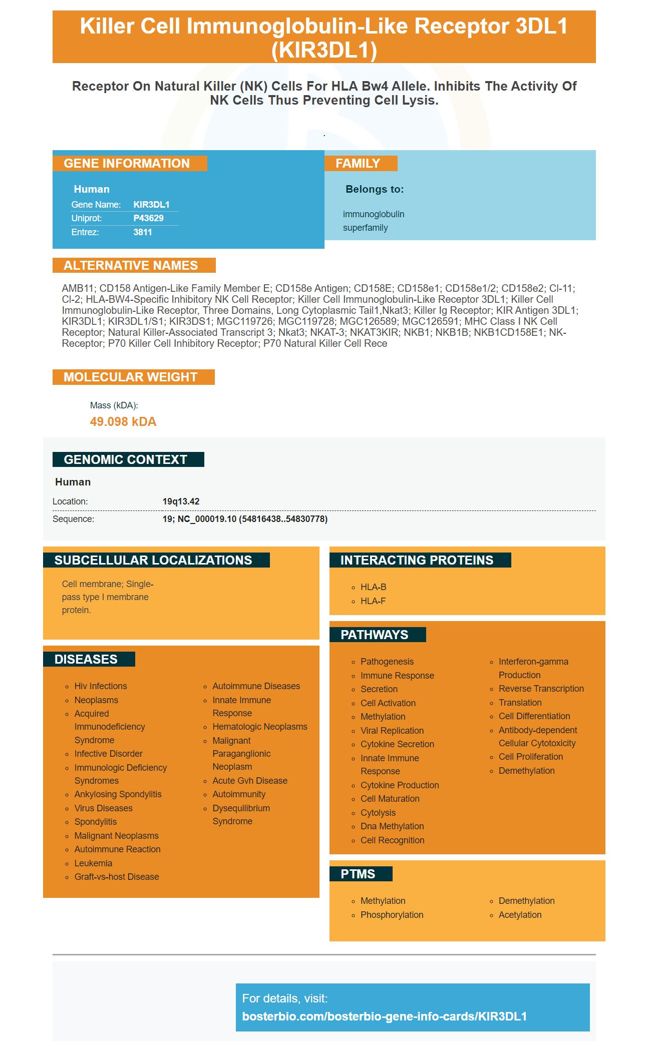
Facts about Killer cell immunoglobulin-like receptor 3DL1.
.
Human | |
---|---|
Gene Name: | KIR3DL1 |
Uniprot: | P43629 |
Entrez: | 3811 |
Belongs to: |
---|
immunoglobulin superfamily |
AMB11; CD158 antigen-like family member E; CD158e antigen; CD158E; CD158e1; CD158e1/2; CD158e2; cl-11; cl-2; HLA-BW4-specific inhibitory NK cell receptor; killer cell immunoglobulin-like receptor 3DL1; killer cell immunoglobulin-like receptor, three domains, long cytoplasmic tail1,nkat3; killer Ig receptor; KIR antigen 3DL1; KIR3DL1; KIR3DL1/S1; KIR3DS1; MGC119726; MGC119728; MGC126589; MGC126591; MHC class I NK cell receptor; Natural killer-associated transcript 3; Nkat3; NKAT-3; NKAT3KIR; NKB1; NKB1B; NKB1CD158E1; NK-receptor; p70 killer cell inhibitory receptor; p70 natural killer cell rece
Mass (kDA):
49.098 kDA
Human | |
---|---|
Location: | 19q13.42 |
Sequence: | 19; NC_000019.10 (54816438..54830778) |
Cell membrane; Single-pass type I membrane protein.
We will be discussing the Anti-KIR3DL1 markinger by Boster Bio as well as its applications in this article. We'll also be discussing the reliability, trustworthiness, and safety of this product. We will also address the cost. We hope you'll find this information useful! Please read this article if you are interested in learning more about KIR3DL1!
Boster bio Anti-KIR3DL1, a commercially available boster antibody is a recombinant, human protein expressed in E.coli. It uses a His–Tag. It is suitable for applications such as immunohistochemistry. The boster antibodies can be stored at -20°C to +8°C for upto one year, or at 4°C to +8°C for upto one month. Each vial of the boster antibody contains four micrograms of Trehalose, 0.9mg NaCl, as well as a peptide that blocks signals from the KIR3DL1 gene.
Boster Bio Anti–KIR3DL1, also known as boster anti–KIR3DL1, an extremely selective monoclonal antibody, induces ADCC (antibody dependent cell cytotoxicity) and is found in significant numbers KIR3DL-expressing cells. The anti-KIR3DL1 marker is designed to bind a protein called chAZ158 and stimulate cell lysis in vitro.
Boster bio anti-KIR3DL1 antibody inhibits the proliferation of KIR3DL1 cells and KIR3DL2 cell. The compounds bind to the common determinant on the KIR3D receptors, preferably the domain D0. They may also stimulate signaling through the KIR3DL protein. The boster bio anti–KIR3DL1 antibody can be purchased in monoclonal or homodimeric form.
Boster Bio Anti–KIR3DLMarker is a useful diagnostic instrument for the detection KIR3DL1 protein expression in human cells. It is highly compatible with KIR3DL1 polypeptides, and can be used to purify and label human cells. It can also assist in diagnosing disease. These antibodies are highly specific to human malignant CD4+ T cells. They can also target specific epitopes within KIR3DL polypeptides. This makes them very useful for medical research.
The bosterbio Anti-KIR3DL1 marker was made with human KIR3DL1 Proteins. Human KIR3DL1 protein is found in NK cell subsets and T lymphocyte subsets. They share similar extracellular areas. The monoclonal antibody for both inhibitory and activating KIR proteins recognises them and are classified according to their sequence analogies.
The antigen-binding chemical inhibits cell proliferation by binding to KIR3DL2 Polypeptide. In addition to inhibiting cell proliferation, it also inhibits ADCC. The antigen-binding compound can be used to inhibit the activity and proliferation of KIR3DL polypeptide-expressing cells in vitro. The antigen-binding compound can also be used to inhibit the growth and proliferation of KIR3DL polypeptide-expressing cells in a variety of different diseases.
KIR3DL1 has been shown useful in analyzing NK cells. KIR3DL1 expression in NK cells helps these immune cells recognize and attack tumors. The marker can also serve to target antigen-presenting (also known to be involved in cancer) cells. KIR3DL1 also has the ability to detect and track viral infections.
KIR3DL1 monoclonal antibody recognizes KIR3DL1 human protein. It does not cross react with other members the KIR family, such as those found in BaF/3 tanning agents. The antibody is provided in a saline solution that contains sodium azide and BSA. Each application will require a different dilution. Our website has general protocols to detect KIR3DL1 protein in human cells.
In animal models, dasatinib improved the degranulation activity in NK cells when they were exposed to KIR3DL1-expressing cells. When compared to KIR3DL1-positive T cells, dasatinib enhanced the degranulation of KIR3DL1+ NK cells against B*52/K562 tumor cells. Dasatinib also enhanced the activity NK cells in response to KIR3DL1-null tumor cells.
Furthermore, the expression of KIR3DL1 in CML patients is associated with a specific pattern of NK cell activation. This novel marker could be used to develop a new therapeutic strategy for increasing anti-tumor NK cells immunity against CML. However, this research is limited due to its unavailability. Further research is needed on how the KIR3DL1 mark affects the antitumor activity CML-derived CML-derivedNK cells.
NK cells are crucial in the immune system. NK cells play a crucial role in the immune system's first line defense against viruses. They also control tumor growth, metastasis, and other aspects of tumor development. HLA-class I specific inhibitory receivers was essential in understanding NK cells' function. Alessandro Moretta, a leading genetic researcher, has done extensive genetic research about KIR3DL1 as well as its polymorphism. The basic concepts behind NK cells function and targeting have been successfully translated into human haploidentical stem-cell transplantation.
KIR3DL1 levels in NK cell phosphorylation are also correlated with ERK1/2 expression. The results also showed that KIR3DL1 was able to induce higher frequencies of CCL4 and IFN-1-g secretion in NK cells. These results suggest that KIR3DL1 -transducedNK cells are capable to stimulate inflammatory reactions.
KIR3DL1 gene marker accuracy and reliability are dependent on their ability to distinguish between different subtypes. Although the KIR3DL1 genetic gene has been identified and used for research since 1996, further testing is needed in order to establish its clinical utility. Genetic testing is currently the most reliable way to determine whether a gene causes disease or not. It is the most preferred method for genetic testing for cancer.
The KIR3DL1 marker does not have a reliable way to determine disease risk, unlike other genetic markers. This marker can however be used to diagnose and treat cancer and other diseases. The underlying mechanism is not clear, and more research is needed. It may be due to in-vivo condition. The KIR3DL1 genetic cluster was only recently identified at high resolution.
The KIR3DL1 gene contains two different alleles. One of the two alleles in the KIR3DL1 gene carries the isoleucine–leucine variant, while the second carries threonine–alanine. The KIR3DL1 gene marker is trusted only if the other is not inherited by a parent. This is especially true for fathers who inherit the other gene.
The KIR3DL1 marker plays a pivotal role in natural killer cell (NK) proliferation. The marker is produced by NK cell as part of a cytotoxic t-cell response. KIR3DL1 has several targets for serine/threonine/kinases. KIR activation results in activation of signaling downstream DAP12 and PCL-g1, Syk/ZAP70, calcium mobilization.
The KIR3DL1 gene is highly conserved among individuals. However, it has recently been linked to a number of diseases, including cancer. The exact nature of the interactions between KIR3DL1 and KIR3DS1 remains a mystery. KIR3DL1 testing is expensive. Before ordering genetic tests for KIR3DL1, it is important to calculate the cost of KIR3DL1.
The KIR3DL1 molecule recognizes a Bw4 epitope located on the C-terminus a subset MHC class I molecules. Polymorphisms of the KIR3DL1 genetic gene could alter the strength or inhibitory potential this interaction. However, small sample sizes can influence specific allele associations. KIR3DL1*00501, KIR3DL1*01301 and KIR3DL1*00501 are also associated with a trend towards BD.
In killing assays of mice, the KIR3DL1+ and KIR3DL1+ subsets were significantly more responsive to Bw4/4 than their counterparts. This may be due in part to differences in the BD or KIR3DL1 protein receptors. Although the KIR3DL1+ is more responsive, evidence is lacking to support a causal connection between the two. To confirm these findings, further studies will be needed on the KIR3DL1+ cells.
PMID: 7716543 by Colonna M., et al. Cloning of immunoglobulin-superfamily members associated with HLA-C and HLA-B recognition by human natural killer cells.
PMID: 8777725 by Wagtmann N., et al. Killer cell inhibitory receptors specific for HLA-C and HLA-B identified by direct binding and by functional transfer.