This website uses cookies to ensure you get the best experience on our website.
- Table of Contents
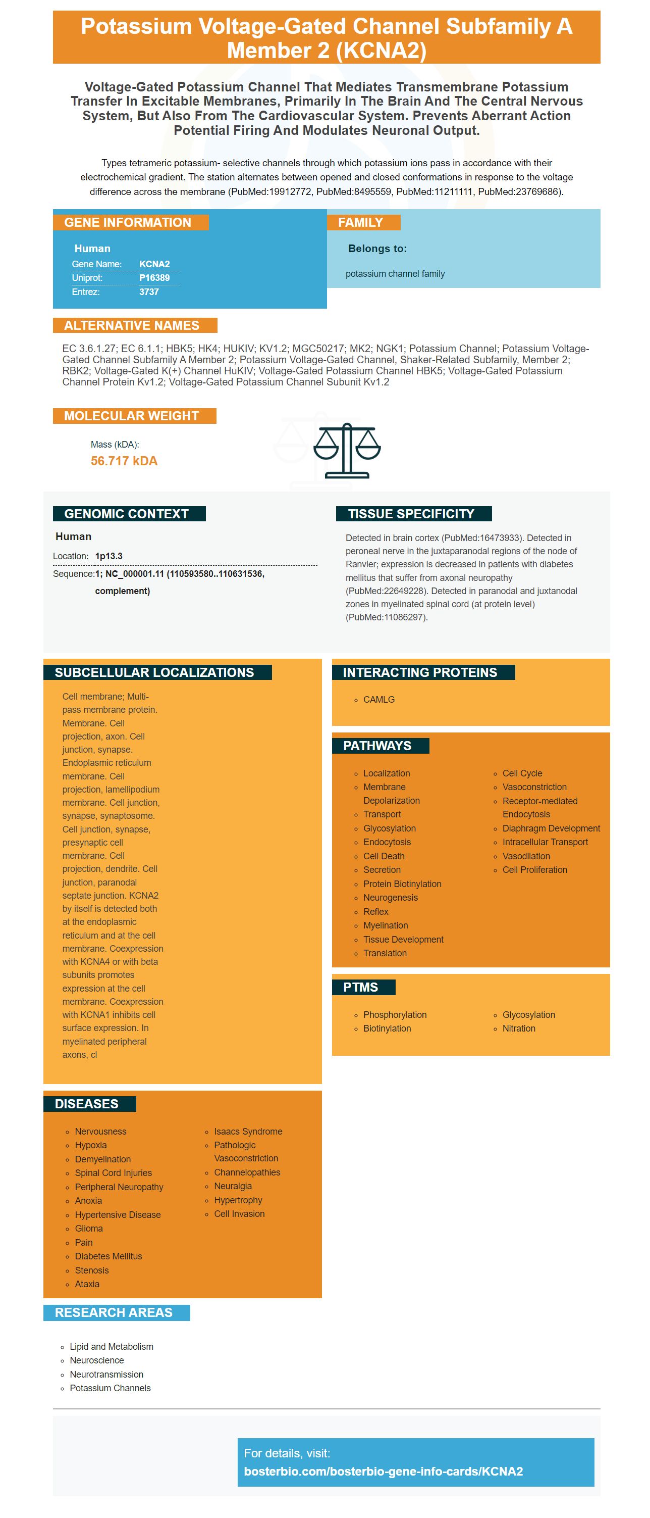
Facts about Potassium voltage-gated channel subfamily A member 2.
Types tetrameric potassium- selective channels through which potassium ions pass in accordance with their electrochemical gradient. The station alternates between opened and closed conformations in response to the voltage difference across the membrane (PubMed:19912772, PubMed:8495559, PubMed:11211111, PubMed:23769686).
Human | |
---|---|
Gene Name: | KCNA2 |
Uniprot: | P16389 |
Entrez: | 3737 |
Belongs to: |
---|
potassium channel family |
EC 3.6.1.27; EC 6.1.1; HBK5; HK4; HUKIV; KV1.2; MGC50217; MK2; NGK1; potassium channel; potassium voltage-gated channel subfamily A member 2; potassium voltage-gated channel, shaker-related subfamily, member 2; RBK2; Voltage-gated K(+) channel HuKIV; Voltage-gated potassium channel HBK5; voltage-gated potassium channel protein Kv1.2; Voltage-gated potassium channel subunit Kv1.2
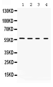
Mass (kDA):
56.717 kDA
Human | |
---|---|
Location: | 1p13.3 |
Sequence: | 1; NC_000001.11 (110593580..110631536, complement) |
Detected in brain cortex (PubMed:16473933). Detected in peroneal nerve in the juxtaparanodal regions of the node of Ranvier; expression is decreased in patients with diabetes mellitus that suffer from axonal neuropathy (PubMed:22649228). Detected in paranodal and juxtanodal zones in myelinated spinal cord (at protein level) (PubMed:11086297).
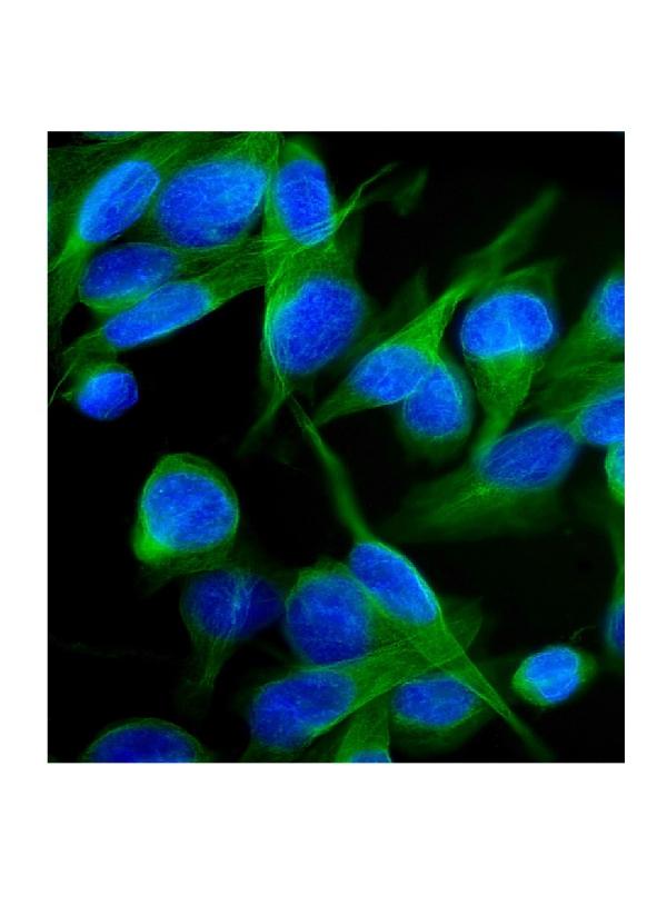
Cell membrane; Multi-pass membrane protein. Membrane. Cell projection, axon. Cell junction, synapse. Endoplasmic reticulum membrane. Cell projection, lamellipodium membrane. Cell junction, synapse, synaptosome. Cell junction, synapse, presynaptic cell membrane. Cell projection, dendrite. Cell junction, paranodal septate junction. KCNA2 by itself is detected both at the endoplasmic reticulum and at the cell membrane. Coexpression with KCNA4 or with beta subunits promotes expression at the cell membrane. Coexpression with KCNA1 inhibits cell surface expression. In myelinated peripheral axons, cl
An immunofluorescence test is used to detect if there is an underlying immune system disorder. The patient had autoantibodies to the KCNA2 KC protein in his serum. However, CSF analysis five months later in 2020 showed no such antibodies. Instead, elevated neuronal specific enolase, tau protein and Ass42/40 were discovered. CSF testing in the patient ruled out Lues as well as neuroborreliosis.
Although KCNA2 autoantibodies were detected in the serum of only two patients, they were found to be related to paraneoplastic diseases. KCNA2 autoantibodies may contribute to cognitive impairment and are related to a specific intracellular epitope. These findings suggest that KCNA2 autoantibodies may be associated with the development of Alzheimer's disease. The clinical significance is not yet clear.
The phenotypic profile of KCNA2 has a wide range. It is not clear if it is involved. The phenotypic profile for KCNA2 mutants is varied and includes a variety inflammatory and degenerative disorders. It is still unclear how KCNA2-AS is involved in the pathogenesis of PHN. The current study aims at identifying the underlying mechanism of KCNA2-AS and their clinical significance.
KCNA2-AS has been found in the spinal cord tissue of PHN-model rats and has been shown that it can alleviate mechanical allodynia. Knockdown of this binding protein reduces pSTAT3 expression in PHN-model rats. Knockdown KCNA2AS inhibits spinal astrocyte stimulation and may be a possible target therapy for PHN.
Antibody detection in the serum was crucial in this case. The immunofluorescence test revealed that the serum contained no specific autoantibodies. However, it showed elevated levels for neuron-specific and tau proteins, which were signs of neuronal damage. Also, the ratio Ass42/40 was lower than expected and ruled out either neuroborreliosis (or Lues).
In addition to identifying an association between the presence of antibodies against KCNA2 and the risk of neurological disease, KCNA2 also plays an important role in the development of neuropathic pain. It is also associated with DNMT3a, which suppresses Kcna2 transcription. It is also related to epileptic encephalopathies and hereditary spastic paraplegia.
Genepharma synthesized biotinylated probes for KCNA2 AS/NC and transfected them into primary rat spinal cord astrocytes. Lysis Buffer was used after 48 hours to harvest the cells. After transfection, RNA was extracted using an RNase Pull-down kit. The sample was then processed for qRT-PCR.
Although there is not a consensus, KCNA2 autoantibody research should be done in patients with cognitive impairment. The studies should include specific cohorts with cerebrospinal fluid as well as laboratory parameters. These studies should specifically identify the epitopes and determine the mode or action of the antibodies. They also need to look for coexisting pathogenetic antibodies.
Anti-ALD antibodies created by patients recognize human Neuroantigens. This suggests that they may be biomarkers and diagnostic markers for neuronal impairment associated with different diseases. Moreover, Dale found that the antibodies developed in patients recognized murine, rather than human neuroantigens, indicating that a murine Ab was a viable alternative to synthetic Ab.
Memory clinics are increasingly using neuronal autoantibody detection. A specific diagnostic approach to neuronal antibodies and therapeutic management is necessary as they increase in frequency. This review aims to summarize the current nosology of neural autoantibody-associated dementia. Literature on the subject was collected using PubMed. It includes a review of clinical and laboratory findings. The authors conclude that there is no specific treatment for patients with these autoantibodies, but that further studies should be performed to establish a definitive diagnosis.
Although anti-neuronal antibody have been associated with epilepsy in some cases, their significance is not known. However, they could be due to tissue destruction. Consequently, immunosuppression may be beneficial in treating epilepsy. It is important that you recognize the clinical features and characteristics of each patient before you start any treatment. This study's results should inform future research. In theory, further studies will determine the best patient population to perform anti-body testing.
These findings are promising, despite the limitations. These antibodies could be used as a diagnostic biomarker for Alzheimer's disease. This study examined epilepsy developed after thirty years of aging, hippocampal swelling, and temporal line sclerosis. The patients with cognitive impairment were more likely have higher levels of these antibodies that those with normal or healthier levels.
Despite its limitations the review found 15 studies that met the inclusion criteria. These studies included ten prospective cohort studies and three control studies. Majoie (2006), Wright 2016 & Verrotti 2003 used similar criteria to evaluate their quality. However, the studies did not receive high marks. Despite their poor quality, these studies did show positive antibody tests for patients with cognitive impairment.
The study revealed that serum anti-TBP levels (KCNQ), were higher among AD patients than those in control subjects. These results indicate that high levels anti-TBP (KCNQ) might be a risk factor for AD, but the significance of these results is still unknown. Researchers also suggested that ethnic composition could be determined by the distribution of skin colour in the population. The study design is not perfect.
This study identifies the first documented case of KCNA2 antibody production in CSF in patients with a paraaneoplastic neurological disease. To determine if patients have antibodies to KCNA2, serum was analyzed and CSF was also examined. A diagnostic biochip array, which included recombinant HEK293 cells, was used to detect the presence of KCNA2 autoantibodies.
To assess the role of the antibody, KCNA2 antibody studies are required. They could be a potential surrogate marker, as they are associated to paraneoplastic neuropathy syndrome. They should be investigated in cognitively impaired patients with a certain paraneoplastic backdrop, and cerebrospinal Fluid samples should also be assessed. Researchers should also seek to determine the epitope location and mechanisms of action of KCNA2 autoantibodies, and to identify any co-existing pathogenetic autoantibodies.
There are differences between the groups, however. In group A, there were six cases of axonal neuropathy, while in group, only one case was identified. Although autoimmunity is strongly implicated in both groups, these cases are different enough to make it difficult to draw a definitive conclusion. These studies have implications in the management of patients with paraneoplastic neural syndrome.
A 54 year-old woman was diagnosed with lung carcinoma. She presented with symptoms of cerebellar and lymph node metastasis. Although anti-N-methyl-d-aspartate receptor antibodies and paraneoplastic syndrome were not detected in this patient, the diagnosis was confirmed. A patient with paraneoplastic neurological disorder was diagnosed with nivolumab in-induced acute cerebellar tremor. The condition resolved within one week.
The study also revealed that patients with anti-KCNA2 antibody had a wide range of pain phenotypes. One patient suffered from erythromelalgia like pain, while two others had severe neuropathy and progressive dementia. The patient with positive autoantibodies also had a history for peripheral vascular diseases. This study also highlights how important it is to understand autoimmune pathophysiology, and the role that autoantibodies play in the treatment for diseases caused by the immune systems.
These results, in addition to KCNA2 antibody studies, also suggest the potential role that the CNS immunogen may play in the treatment for SN. Cerebellar atrophy, which is a rare condition, may be a contributing factor to the symptoms of SN, especially ataxia. In addition to this, the clinical presentation of the syndrome may indicate a diagnosis of paraneoplastic neurological syndrome.
A wide range of therapeutic options have been opened up by the classification of PNH. The immune system is the most common mechanism of the disease. This includes antibodies against voltage-gated sodium channel (VGKC). Other types are caused toxins and genetic disorders. Patients with paraneoplastic neurological diseases are more likely to detect VGKC antigens.
PMID: 19912772 by Ramaswami M., et al. Human potassium channel genes: molecular cloning and functional expression.
PMID: 8495559 by Po S., et al. Heteromultimeric assembly of human potassium channels. Molecular basis of a transient outward current?