This website uses cookies to ensure you get the best experience on our website.
- Table of Contents
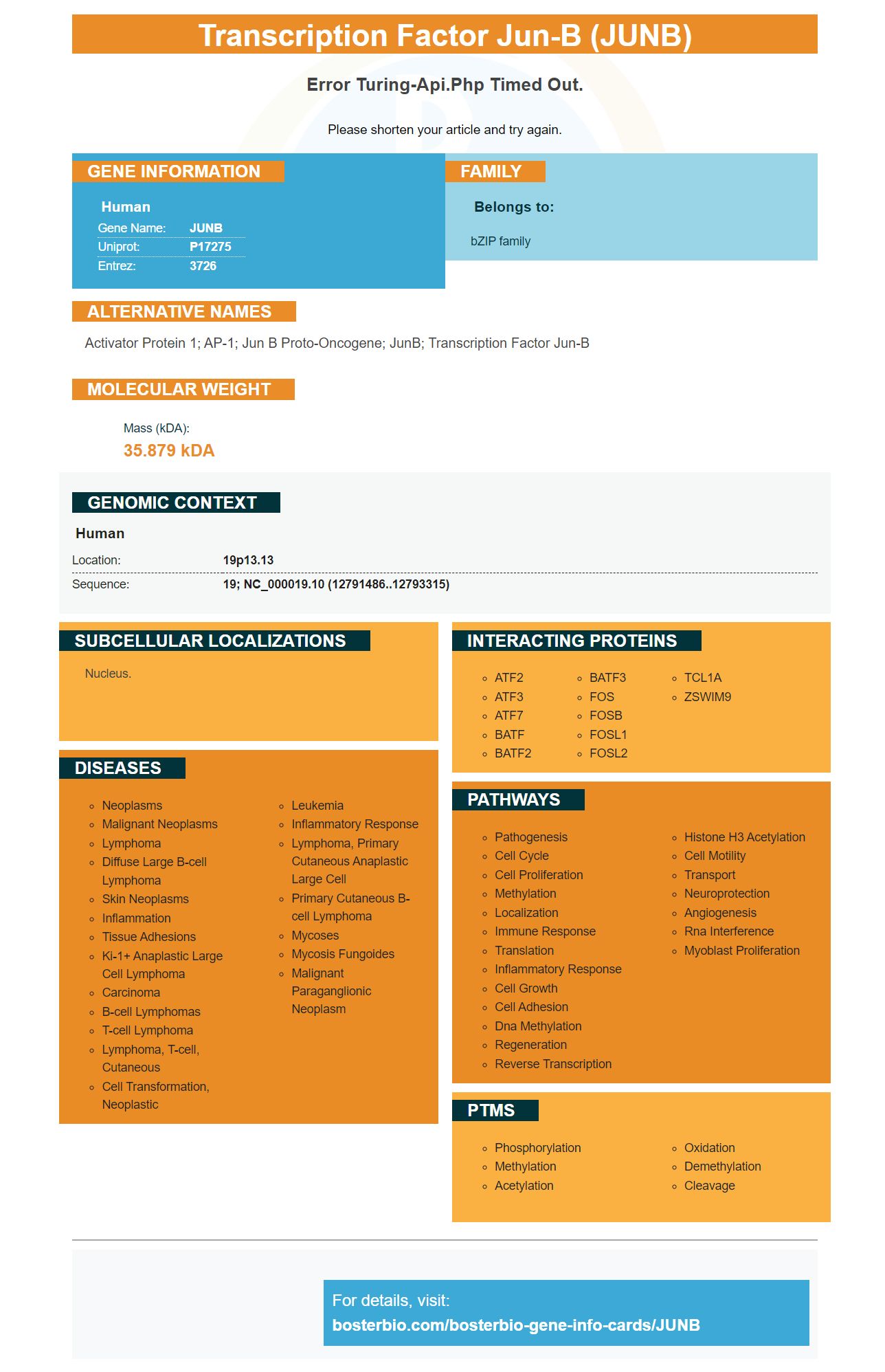
Facts about Transcription factor jun-B.
Please shorten your article and try again.
Human | |
---|---|
Gene Name: | JUNB |
Uniprot: | P17275 |
Entrez: | 3726 |
Belongs to: |
---|
bZIP family |
activator protein 1; AP-1; jun B proto-oncogene; JunB; transcription factor jun-B
Mass (kDA):
35.879 kDA
Human | |
---|---|
Location: | 19p13.13 |
Sequence: | 19; NC_000019.10 (12791486..12793315) |
Nucleus.
If you're looking for a monoclonal antibody that targets the JUNB marker, you've come to the right place. We'll discuss the best uses of this antibody in the context of obesity research and how it differs from other anti-obesity antibodies. In this article, we'll also discuss how to optimize the performance of experiments with Boster Bio.
The Boster Bio Anti-JUNB Monoclonal is a highly validated antibody designed specifically for flow cytometry. These monoclonal antibodies are produced in-house and are derived from well-established clones. They are available conjugated for ICC/IF, WB, ChIP, and IHC applications. During the checkout process, the customer must select the conjugate desired.
The antiserum is purified by immunoaffinity chromatography (IAC) to remove rabbit serum proteins. The antiserum is 100% pure. It is then diluted in 0.01 M PBS with 0.01% Thimerosal. This working solution must be optimized for the end use. The buffer can be stored for a long period of time by adding 1% sodium acetate or 0.05% NaN3.
Neutralization studies showed that all nine monoclonal antibodies tested were active against Wu01, Delta, and Beta strains. Seven out of nine showed neutralizing activity against Omicron variant, but the activity was reduced or abolished in one of them. This reduced neutralizing activity may limit treatment options against COVID-19 induced by Omicron. It should be noted that the anti-JUNB Monoclonal Antibody from Boster Bio was also evaluated against SARS-CoV-2.
This mouse-based monoclonal antibody to adiponectin was validated in human tissue in IHC studies. It contains 100ug of antigen per vial and is ideal for detecting adiponectin in fat tissue. The product is suitable for research purposes and can be purchased from Boster Bio. For more information, please visit their website.
Human adipose tissue was incubated with three mAbs. They all stained the tissue, although the staining patterns of the three antibodies were not significantly different. All three mAbs recognized adiponectin in the nuclei of adipocytes and in blood vessels. Similarly, the three mAbs showed differential staining of other human tissues. KH7-41 and KH7-33 antibodies stained lung tissues, while KH4-8 mAb recognized both MMW and HMW isoforms.
Boster Bio uses Cas9 proteins to produce monoclonal antibodies to over 20,000 unique targets. These antibodies are suitable for Western blotting. They can also detect protein-protein interactions with Cas9-expressed cell lines. They use biotin-conjugated antibodies to detect adiponectin in human tissues.
The development of therapeutic antibodies for adiponectin mAbs is underway. The aim is to target detrimental isoforms of adiponectin and keep the beneficial forms intact. This will allow researchers to develop adiponectin antibodies for human diseases. The antibodies are a vital component of many research studies. Once approved, they could be used as effective treatments for various diseases.
The mAbs for adiponectin were generated in hybridomas. These mAbs were validated in ELISA and Western blot assays. The IgGs were purified from the culture supernats using a Protein G column. The purity of the purified IgG was determined using Coomassie blue staining. The mAbs were then probed with an anti-mouse IgG secondary antibody coupled with horseradish peroxidase.
The mAbs also inhibited pro-inflammatory cytokines in CIA mice. In addition, histology of CIA mice knee joints demonstrated that they inhibited inflammatory processes. mAbs that recognize adiponectin's epitope, called KH4-8, inhibited the development of arthritic conditions in CIA mice.
The JUNB marker has several potential applications for research. It is a glycoprotein that is expressed in the plasma as dimeric and multimeric forms, and also on the cell surface and in the extracellular matrix. It is an important component of cell adhesion and migration processes. Cellular fibronectin differs from plasma fibronectin in that it affects the morphology of transformed cells and their hemaglutination. The antibodies manufactured by Boster Bio have been validated on multiple platforms to maximize their potential use.
PMID: 2513129 by Schuette J., et al. jun-B inhibits and c-fos stimulates the transforming and trans- activating activities of c-jun.
PMID: 2112242 by Nomura N., et al. Isolation of human cDNA clones of jun-related genes, jun-B and jun- D.