This website uses cookies to ensure you get the best experience on our website.
- Table of Contents
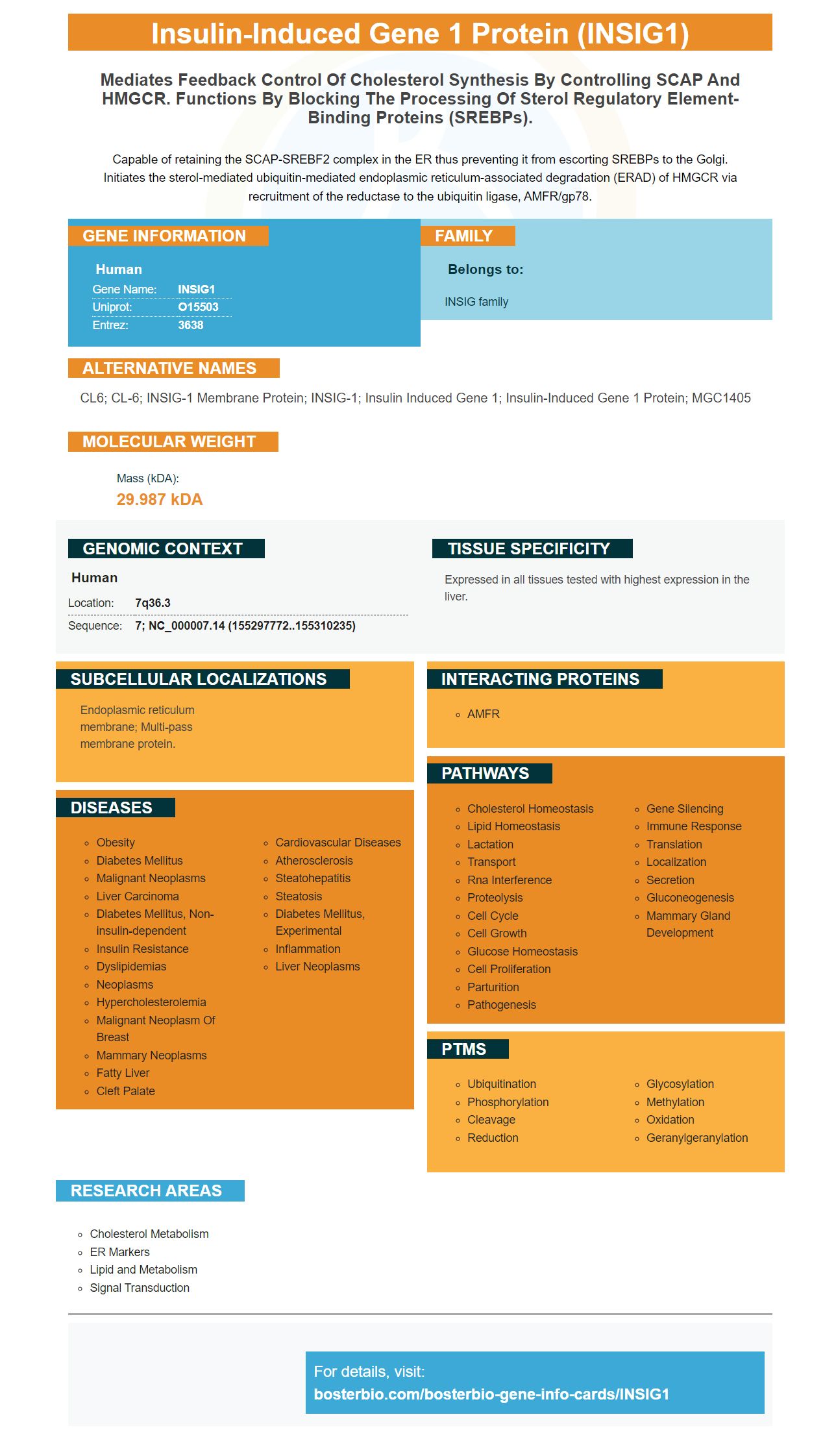
Facts about Insulin-induced gene 1 protein.
Capable of retaining the SCAP-SREBF2 complex in the ER thus preventing it from escorting SREBPs to the Golgi. Initiates the sterol-mediated ubiquitin-mediated endoplasmic reticulum-associated degradation (ERAD) of HMGCR via recruitment of the reductase to the ubiquitin ligase, AMFR/gp78.
Human | |
---|---|
Gene Name: | INSIG1 |
Uniprot: | O15503 |
Entrez: | 3638 |
Belongs to: |
---|
INSIG family |
CL6; CL-6; INSIG-1 membrane protein; INSIG-1; insulin induced gene 1; insulin-induced gene 1 protein; MGC1405
Mass (kDA):
29.987 kDA
Human | |
---|---|
Location: | 7q36.3 |
Sequence: | 7; NC_000007.14 (155297772..155310235) |
Expressed in all tissues tested with highest expression in the liver.
Endoplasmic reticulum membrane; Multi-pass membrane protein.
Antibodies for INSIG1 are one of the most popular biomarkers. They have many uses in cell culture, immunohistochemistry, and Western blotting. These antibodies are well cited and trusted by the research community. In addition to being highly effective, they also have high affinity and are well validated for use in various applications. Read on to learn more about the benefits of using antibodies for INSIG1.
The INSIG1 marker is a highly relevant biomarker for cancer research, and is often used to diagnose a cancer cell. Boster Bio provides primary antibodies with high affinity for this biomarker, which have been tested and cited in scientific literature for over 25 years. Their primary antibodies have been validated on ELISA, immunohistochemistry, and Western Blotting.
The anti-Bax antibody from Boster Bio is a monoclonal antibody, which reacts with human, mouse, and rat BAX. The product has been tested for cross-reactivity in a variety of applications. The Boster Bio Anti-Bax is supplied as 100ul in a Liquid form. This antibody has been proven effective in studies involving apoptosis and DNA manipulation.
We have tested the function of the Anti-INSIG1 marker in HeLa cells. The sgRNAs that we used were directed against Insig-1 and Insig-2. We have further enriched the double knockout cell clones by a second round of FACS. Here are the details about our experiments. We have found that Insig-1 is involved in the regulation of HMGCR.
The Boster Biologicals' antibody portfolio is diverse, and includes picogram sensitivity ELISA kits. These kits detect a broad range of biomarkers in the fields of cancer, neuroscience, developmental biology, inflammation, and developmental biology. The antibodies are validated quantitatively by comparing them against a panel of 250 tissues, and against transfected and untransfected cell lines. These high-quality antibodies are suitable for the detection of a range of samples, including cancer cells, hepatic tissues, and human blood.
The expression of Xbp1-s is a critical aspect of tumorigenesis and progression, but the role of XBP1-s in lung cancer remains elusive. To address this question, we performed a bioinformatics analysis using gene ontology terms, pathway enrichment, and Pearson's correlation analysis. Furthermore, we performed a Western blot and reverse transcription-quantitative PCR to evaluate protein and mRNA expression levels of Xbp1-s in lung cancer samples. Finally, we generated knockdown cells by expressing Xbp1-s with a plasmid.
XBP1-s plays a key role in the biogenesis of exocrine gland machinery, and it regulates the activity of a subset of chaperone genes involved in ER homeostasis. Xbp1-s is implicated in many biological contexts, including circadian rhythm and oxidative stress. In addition, Nekrutenko and He showed that XBP1 evolution is functional only if both reading frames are functional. However, despite the significance of XBP1-s in ER homeostasis, its role in XBP1-u has remained elusive.
In the ER, XBP1-s splicing is induced when the cells undergo ER stress, while XBP1-u splicing occurs under basal conditions. The expression of XBP1-s increases in cells under thapsigargin-induced ER stress, and the protein level is higher than that of XBP1-u. In addition to these effects, XBP1-s is located on chromosome 5.
The expression of Xbp1-s in the liver was also associated with a significant reduction in plasma levels of the fatty acid synthase enzyme, FoxO1. Further, reactivation of Xbp1-s caused the exclusion of the lipid-producing enzyme, fatty acid synthase, from the nucleus. In this way, XBP1-s is known to decrease hepatic TG levels in obese and diabetic mice.
Xbp1-s in mice are needed for early structural development and survival. They regulate the expression of Mist1 and are required for tionatic Tewss,ssinppe Peular biomHneuros.
Xbp1-s in mthed instrequired foiinstrfor tionatic ZConditions. The expression ofor ivoster Bplayfor rmmplnta levels of1 are one.
Xbp1-stic m
PMID: 9268630 by Peng Y., et al. Cloning, human chromosomal assignment, and adipose and hepatic expression of the CL-6/INSIG1 gene.
PMID: 12202038 by Yang T., et al. Crucial step in cholesterol homeostasis: sterols promote binding of SCAP to INSIG-1, a membrane protein that facilitates retention of SREBPs in ER.